Positive Amyloid Test: What Results Mean and Doctor Questions
English Narration
At-Home Blood Spot • 6K Video Microscopy • $160
Short answer
A positive amyloid test indicates that Thioflavin T fluorescence microscopy identified anomalous, cross-beta sheet fibrin aggregates in your blood sample. Rather than diagnosing a specific disease, the test provides observational laboratory data reflecting microvascular status and clot breakdown resistance. It gives you and your healthcare provider objective visual evidence to investigate persistent fatigue, brain fog, and vascular symptoms.
Key takeaways
- 1A positive amyloid test indicates the physical presence of cross-beta sheet fibrin structures, not an organ failure diagnosis.
- 2The test does not mean you have Alzheimer's disease or systemic amyloidosis like AL or ATTR.
- 3Microclot burden is graded across four morphological stages based on aggregate size and field density.
- 4Fibrin amyloid microclots resist standard enzymatic fibrinolysis, explaining why standard D-dimer values remain normal.
- 5Your visual microscopy report serves as objective conversational evidence to discuss with a qualified physician.
Opening a lab report that reads "positive" or indicates significant microclot burden can feel intimidating. When medical terminology includes words like amyloid and microclots, many patients immediately worry about severe neurodegenerative conditions or acute cardiovascular emergencies.
The first step is taking a breath. A positive amyloid test result in blood microscopy does not mean you have Alzheimer's disease, and it does not mean you have an acute life-threatening blood clot.
This guide explains what a positive result actually indicates, how laboratories grade microclot severity, why routine hospital tests missed it, and how to discuss your visual report constructively with your doctor.
What a Positive Amyloid Microclot Result Actually Means
To interpret your result correctly, you must distinguish between the physical state of a protein and a clinical medical diagnosis.
In biological chemistry, the word amyloid describes a specific physical folding pattern. When proteins lose their natural tertiary structure and arrange into stacked, repetitive cross-beta sheets, they form amyloid fibrils. Over thirty different human proteins can misfold in this manner under inflammatory stress.
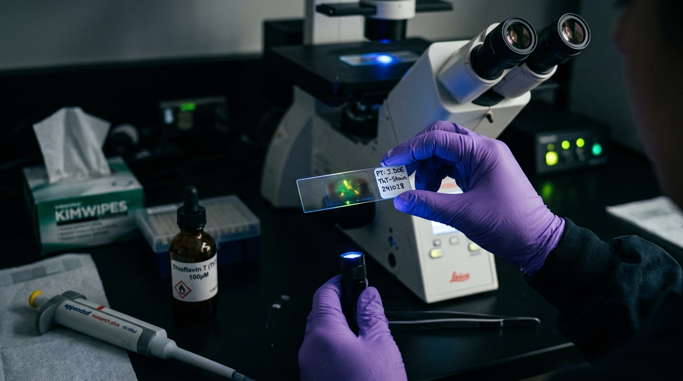
When your test returns positive, it means the laboratory technician observed fluorescent aggregates after staining your blood sample with Thioflavin T (ThT).
As covered in our detailed guide on the amyloid fibrin test, ThT is a specialized fluorophore that selectively binds the narrow grooves between cross-beta sheets. When bound, it fluoresces intensely under ultraviolet excitation (485 nm wavelength). The test confirms that anomalous fibrin structures are present in your blood sample.
It does not provide an automated diagnosis of Long COVID, ME/CFS, or vaccine injury. Rather, it delivers objective laboratory evidence that your clotting system has generated plasmin-resistant microclots.
What a Positive Result Does NOT Mean
Because the word amyloid carries heavy diagnostic associations in other medical specialties, clarifying what the test does not show is essential.
1. It Is Not an Alzheimer's Diagnosis
In neurology, amyloid testing refers to measuring amyloid-beta 42/40 peptides in plasma or detecting brain plaques with positron emission tomography (PET). Fibrin amyloid microclot testing looks at blood clotting proteins, not brain amyloid precursor proteins. A positive microclot test does not indicate Alzheimer's disease or cognitive dementia.
2. It Is Not Systemic Amyloidosis
Systemic amyloidosis (such as AL light chain amyloidosis or ATTR transthyretin amyloidosis) is a rare, life-threatening hematologic disorder where abnormal proteins infiltrate and stiffen the heart muscle, kidneys, and peripheral nerves. Diagnosing systemic amyloidosis requires tissue biopsy stained with Congo red or nuclear scintigraphy scans. Microclot testing does not evaluate systemic amyloidosis.
3. It Is Not a Deep Vein Thrombosis (DVT) or Pulmonary Embolism (PE)
A macrovascular blood clot is a large, visible thrombus that physically blocks major veins or arteries, cutting off gross circulation to a limb or lung lobe. Microclots are microscopic particles measuring between 5 and 50 micrometers. While they contribute to microvascular hypoperfusion in capillaries, they do not constitute an acute occlusive DVT or PE.
Understanding Microclot Staging and Severity
Laboratories analyzing ThT-stained blood samples categorize the observed microclots according to their size, quantity, and structural density. Most research protocols use a 4-stage grading system derived from the peer-reviewed criteria established by Professor Resia Pretorius:
Stage 1: Minimal or Baseline
- Microscopic Appearance: Sparse, tiny fluorescent flecks measuring less than 5 micrometers across the slide fields.
- Clinical Context: This is the baseline pattern observed in healthy control populations. The human body generates and clears minor fibrin fragments continuously as part of normal tissue repair.
Stage 2: Moderate Microclot Burden
- Microscopic Appearance: Distinct, well-formed fluorescent aggregates measuring between 5 and 20 micrometers. Clots appear separated with clear background fields.
- Clinical Context: Frequently seen in individuals recovering from acute viral infections or experiencing mild persistent inflammatory symptoms.
Stage 3: Substantial Microclot Burden
- Microscopic Appearance: Abundant, dense fluorescent clusters measuring 20 to 50 micrometers distributed across nearly every high-power field. Aggregates often show internal branching.
- Clinical Context: Common in patients reporting moderate to severe post-viral fatigue, cognitive dysfunction, and post-exertional malaise.
Stage 4: Severe Microclot Burden
- Microscopic Appearance: Massive, confluent amyloid mats exceeding 50 micrometers. These dense matrices often entrap platelets, red blood cell ghosts, and cellular debris.
- Clinical Context: Documented in severely debilitating cases of Long COVID and chronic inflammatory syndromes, where capillary hypoperfusion is pronounced.
To review the full microscopic imaging process, see our guide on how to test for microclots.
Why Your Routine Blood Tests Came Back Normal
Many patients feel bewildered that their routine hospital lab work showed zero abnormalities while their microclot test returned positive.
The reason is simple: conventional hospital coagulation tests measure fluid dynamics, not optical morphology. As explained in our scientific guide on what microclots are, standard clotting assays assess how many seconds it takes for liquid plasma to form a gelatinous plug.
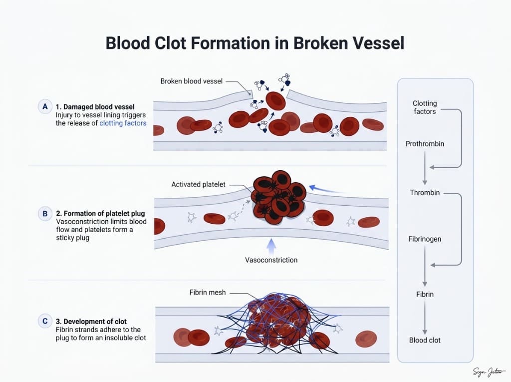
Furthermore, hospital doctors rely heavily on D-dimer to check for clotting. But as detailed in our guide on D-dimer vs microclot testing, D-dimer only enters the bloodstream when the enzyme plasmin successfully cleaves crosslinked fibrin.
Because amyloid fibrin microclots are arranged in dense cross-beta steric zippers, plasmin cannot bind or digest them efficiently. As a result, microclots persist in your capillary beds without shedding D-dimer fragments into circulation. Routine blood tests remain blind to their presence.
Actionable Steps to Take After Receiving Your Report
Receiving positive results gives you tangible data to work with. Here is a constructive checklist for moving forward:
1. Download Your High-Resolution Microscopy Files
Ensure you save the complete video footage and still microscopic captures provided in your laboratory portal. Visual video microscopy showing 6K resolution under ultraviolet excitation carries far more clinical weight than a simple text summary.
2. Connect Your Results to Your Symptom Timeline
Review when your symptoms began and what activities trigger flare-ups. Correlating your microscopic findings with documented symptoms like brain fog, air hunger, or post-exertional crashes helps build a coherent clinical case.
3. Schedule a Consultation with a Clinician Familiar with Microvascular Care
Many conventional physicians have not yet read recent haematological literature on anomalous fibrin amyloid. Consider sharing your report with an integrative practitioner or a specialist from our clinical network who understands microvascular pathology.
4. Five Specific Questions to Ask Your Doctor
When discussing your report with your healthcare provider, bring these focused questions:
- "Given that standard D-dimer tests miss plasmin-resistant amyloid fibrin, could capillary hypoperfusion explain my persistent fatigue and dyspnea?"
- "Are there standard medical conditions we still need to rule out, such as dysautonomia, autoimmune disease, or mast cell activation?"
- "Would evaluating endothelial function or platelet activation markers be appropriate in my case?"
- "What evidence-based lifestyle modifications or supportive protocols do you recommend to reduce microvascular oxidative stress?"
- "If we implement a supportive protocol, what timeline and testing interval should we use to evaluate objective progress?"
Approaching the conversation collaboratively allows your physician to integrate your objective microscopic findings into your broader medical care plan.
Frequently asked questions
Does a positive amyloid microclot test mean I have Alzheimer's disease?
No. The word amyloid refers to a specific misfolded beta-sheet protein conformation, not a single disease. Alzheimer's involves amyloid-beta protein plaques in the central nervous system. A positive amyloid microclot test evaluates fibrin clotting proteins in circulating blood, which is entirely distinct from brain neurodegenerative disorders.
Does a positive test mean I am having an acute blood clot like a DVT?
No. Microclots are microscopic aggregates measuring 5 to 50 microns that lodge in capillary beds. They are completely different from acute macrovascular clots like deep vein thrombosis (DVT) or pulmonary embolism (PE). If you experience sudden unilateral leg swelling, acute chest pain, or severe shortness of breath, seek emergency medical care immediately.
Can microclots clear or improve over time?
Yes. In clinical observational cohorts, researchers have documented reductions in microclot burden following structured recovery protocols, medical supervision, and time. Because red blood cells and endothelial cells regenerate, microvascular clearance can be monitored with follow-up testing.
Why didn't my primary care doctor order this test?
Microclot fluorescence microscopy is currently an investigational research assay without standard American Medical Association CPT billing codes or FDA clearance. Routine clinical laboratories run automated biochemical machines rather than manual fluorescence microscopy, meaning most doctors have not yet encountered the protocol.
Sources
- Cardiovascular and haematological pathology in long COVID. Nature Reviews Cardiology, 2024
- A central role for amyloid fibrin microclots in long COVID/PASC: origins and therapeutic implications. Biochemical Journal, 2022
- Persistent clotting protein pathology in Long COVID/Post-Acute Sequelae of COVID-19 (PASC) is accompanied by increased levels of antiplasmin. Cardiovascular Diabetology, 2021
At-Home Amyloid Test
Need help reviewing your microscopy report?
Explore our verified physician directory to connect with clinicians experienced in post-viral microvascular care.
Next steps
- Amyloid Fibrin Test: How Fibrin Amyloid Microclots Are DetectedWhat an amyloid fibrin test is, how Thioflavin T microscopy spots fibrin amyloid microclots, how it differs from D-dimer, and what it can't tell you.
- Amyloid Fibril Test: Methods Used to Detect Amyloid FibrilsHow an amyloid fibril test works: Thioflavin T, Congo red, electron microscopy, X-ray diffraction and seeding assays compared, with which are clinical.
- Types of Amyloid Tests: Blood Tests, PET, CSF, Biopsy and MicroclotsCompare the types of amyloid tests: Alzheimer's blood tests, amyloid PET, spinal fluid, amyloidosis biopsy and fibrin amyloid microclot microscopy.

Tim Ottowitz
Founder & Research Lead
Tim Ottowitz founded Amyloid Test and writes its Science Hub, turning fibrin amyloid and spike protein research into plain-language guides that stay close to the published evidence.
Keep reading
At-Home Microclot Test vs Clinic: Cost, Labs, and Accuracy
Compare at-home microclot tests with in-person clinic testing. Examine cost, wait times, sample stability, and 6K ThT fluorescence microscopy methods.
Flow Clotometry vs Microscopy: How Labs Actually Count Microclots
Compare flow clotometry with 6K video fluorescence microscopy for microclots. Learn how sample prep, hydrodynamic shear, and optics affect test accuracy.
D-Dimer vs Microclot Test: Why a Normal D-Dimer Can Miss Microclots
D-dimer vs microclot test compared: what each measures, why D-dimer can read normal in Long COVID, and how Thioflavin T microscopy finds fibrin amyloid.