At-Home Microclot Test vs Clinic: Cost, Labs, and Accuracy
English Narration
At-Home Blood Spot • 6K Video Microscopy • $160
Short answer
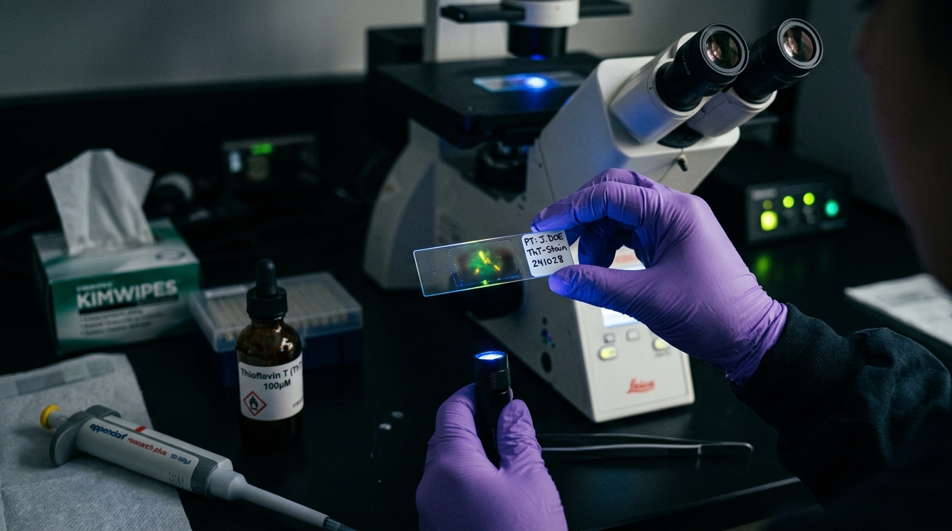
In-clinic microclot testing typically costs $800 to $1,500 and requires traveling to specialized facilities with multi-month waitlists. At-home microclot testing uses a finger-prick blood spot dried on glass, mailed to a dedicated laboratory for Thioflavin T fluorescence microscopy. For $160, patients receive research-grade visual microscopy documentation without cross-country travel, enabling informed discussions with their treating physician.
Key takeaways
- 1Standard hospital coagulation tests like D-dimer miss amyloid microclots entirely because cross-beta sheets resist fibrinolysis.
- 2In-clinic microclot testing often costs $800 to $1,500 and demands long-distance travel to rare specialized centers.
- 3At-home finger-prick collection cards stabilize fibrin amyloid structures, eliminating the rapid platelet degradation seen in uncentrifuged liquid tubes.
- 4A reputable microclot test provides high-resolution 6K microscopy video showing actual Thioflavin T fluorescence, not an arbitrary numerical score.
- 5Neither in-clinic nor at-home microclot tests are FDA-cleared diagnostic devices; both serve as investigational and educational tools.
Patients living with post-viral illness, persistent fatigue, and vascular symptoms frequently hit a brick wall with routine medical testing. Standard blood panels, D-dimer tests, and routine coagulation screens regularly return completely normal results.
This diagnostic blind spot occurs because standard coagulation assays assess soluble clotting factors, not insoluble, amyloid-altered fibrin deposits. When investigating persistent microvascular issues, patients encounter two paths: traveling to a regional clinic or ordering an at-home collection kit.
This buying guide breaks down microclot test costs, logistical differences, sample stability, and optical verification to help you decide which testing route fits your health circumstances and budget.
Why Standard Blood Tests Miss Microclots
Routine hospital hematology relies on biochemical assays developed decades ago. A standard D-dimer test measures circulating protein fragments produced when plasmin breaks down ordinary blood clots.
In post-viral syndromes and inflammatory disorders, fibrin undergoes an anomalous conformational change. As detailed in our guide on D-dimer vs microclot testing, molecules like SARS-CoV-2 spike protein induce fibrinogen to misfold into dense cross-beta sheet structures.
These cross-beta sheets resist enzymatic degradation by plasmin. Because the clots do not break down normally, they generate negligible D-dimer fragments while lingering in capillary beds. Detecting them requires direct optical visualization under fluorescence microscopy using amyloid-binding fluorophores like Thioflavin T (ThT).
In-Clinic Microclot Testing: Overview and Costs
During the initial years of microclot research, testing was available exclusively through academic hospital centers and a small handful of pioneering private practices.
Clinic Availability and Geographic Bottlenecks
In the United States and Europe, only a fraction of specialty practices offer microclot evaluations. Facilities such as MedHelp Clinics in Alabama pioneered clinical microclot processing in North America, drawing patients from across the continent.
Because few commercial laboratories operate the necessary fluorescence microscopes and trained slide preparation technicians, access remains constrained. Patients routinely face 2 to 5-month waiting lists to secure an in-person intake appointment.
Total Out-of-Pocket Expense Breakdown
Because microclot assays lack FDA clearance and AMA billing codes, private health insurance does not cover the service. An in-person testing journey involves several distinct expenses:
- Initial Clinical Intake: Specialized integrative or post-viral medicine clinics charge between $350 and $600 for a new patient evaluation.
- Venipuncture and Centrifugation: Drawing citrated blood and spinning platelet-poor plasma costs approximately $75 to $150.
- Fluorescence Slide Staining: Private laboratory partner fees for ThT staining and manual scoring range from $400 to $750.
- Travel and Lodging: For patients unable to drive locally, flights, hotels, and ground transportation add $500 to $1,500.
In total, obtaining a clinic-based microclot assessment typically requires a total expenditure between $800 and $2,500. For individuals dealing with severe post-exertional malaise or orthostatic intolerance, the physical exertion of long-distance travel presents a significant barrier.
At-Home Microclot Testing: How It Operates
At-home microclot testing was developed to remove geographic barriers and reduce patient costs while preserving the underlying scientific methodology.
Instead of requiring an in-person phlebotomist, an at-home kit utilizes a capillary blood sample collected via an automated lancet. The sample is prepared directly on collection substrates, allowed to dry, and packaged into a protective mailer.
Our research-grade at-home microclot test costs $160 USD (3,200 MXN) with prepaid international shipping to our dedicated imaging laboratory in Mexico City.
The Sample Stability Dilemma: Liquid Plasma vs Dried Blood Spots
The most common technical question regarding at-home testing is sample preservation. In a clinic, phlebotomists draw liquid blood into sodium citrate tubes. If liquid blood sits at room temperature for more than a few hours, platelets undergo spontaneous activation, releasing granules and creating artificial clotting artifacts.
Liquid samples sent across states require precise centrifugation within two hours of phlebotomy, followed by cold packs and expedited courier delivery. When delays occur, sample hemolysis and ex-vivo clotting can invalidate the microscopic scoring.
Dried blood sample collection bypasses this fragility. Once capillary blood is drawn onto a dedicated slide substrate and air-dried, biological enzymatic activity ceases immediately.
Amyloid fibrils possess extraordinary thermodynamic stability. Because cross-beta steric zippers resist temperature fluctuations, enzymatic digestion, and ambient oxidation, dried amyloid structures remain intact during transit. The receiving laboratory then rehydrates and stains the specimen with calibrated Thioflavin T without introducing artificial shear stress.
Side-by-Side Comparison: In-Clinic vs At-Home
The following breakdown summarizes the primary differences between clinic-based evaluation and mail-in testing:
| Feature | In-Clinic Evaluation | At-Home Test Kit ($160 USD) |
|---|---|---|
| Total Cash Outlay | $800 to $2,500 (incl. consults) | $160 USD (all inclusive) |
| Travel Requirement | Mandatory travel to regional hub | Zero travel; completed from home |
| Wait Time for Testing | 2 to 5 months for clinic intake | Kits ship immediately; results in 2–3 weeks |
| Sample Type | Citrated venous blood (phlebotomy) | Capillary blood spot (finger-prick lancet) |
| Sample Degradation Risk | High if shipping liquid tubes; low if run onsite | Low; dried cross-beta sheets remain stable |
| Imaging Resolution | Varies by clinic (often static photos) | 6K RAW 12-bit video microscopy at 20x |
| Clinical Prescription | Often required for laboratory order | No doctor order required to obtain report |
| Medical Guidance | Direct in-person physician interpretation | Objective lab report for your existing physician |
To review registered clinical sites worldwide, consult our directory of microclot testing labs and treatment centers.
What to Look for in a Quality Microclot Test Report
Whether you choose a local clinic or an at-home service, ensure the laboratory delivers objective evidence rather than subjective summaries.
1. High-Resolution Visual Evidence
Microclot analysis should not be reduced to an opaque number on a single sheet of paper. High-quality testing protocols record digital microscopy across multiple slide fields.
Our partner laboratory captures 6K 12-bit RAW video across a 20x optical field under ultraviolet excitation (485 nm excitation, 535 nm emission). Patients receive direct access to their high-resolution footage and still captures, allowing their clinical team to observe actual fibril morphology.
2. Dual Fluorophore Discrimination
Advanced academic research uses multiple fluorophores to distinguish different clot components. Thioflavin T stains the cross-beta amyloid backbone of fibrin. Platelet markers like CD62P (P-selectin antibodies) identify activated platelet aggregates. A comprehensive report distinguishes whether microclots are dense fibrin sheets, loose platelet conglomerates, or mixed structures.
3. Clear Staging Methodology
Laboratories typically evaluate microclots across four stages based on the classification models established by Professor Resia Pretorius and colleagues:
- Stage 1 (Normal / Minimal): Small, sparse fluorescent specks measuring under 5 microns. Typical for healthy individuals without chronic symptoms.
- Stage 2 (Moderate): Discrete microclots measuring between 5 and 20 microns with observable amyloid fluorescence.
- Stage 3 (Substantial): Numerous aggregated microclots measuring 20 to 50 microns across multiple fields of view.
- Stage 4 (Severe): Massive, confluent amyloid mats exceeding 50 microns that actively entrap platelets and cellular debris.
Understanding these stages provides a reliable baseline if you and your doctor decide to monitor changes over time.
Differentiating Microclots from Other Amyloid Conditions
Before ordering any evaluation, ensure you are ordering the appropriate test for your clinical questions. As detailed in our breakdown of the types of amyloid tests, the term amyloid applies to multiple distinct biomedical fields.
An at-home microclot test checks solely for microvascular fibrin amyloid aggregates associated with inflammatory and post-viral conditions. It does not evaluate systemic organ amyloidosis:
- Not an Alzheimer's Test: Does not measure cerebral amyloid-beta 42/40 ratios or phosphorylated tau.
- Not a Systemic Amyloidosis Screen: Does not evaluate immunoglobulin light chain (AL) or transthyretin (ATTR) amyloidosis.
- Not an Acute DVT Rule-Out: Does not replace hospital venous duplex ultrasound for deep vein thrombosis or pulmonary embolism.
How to Discuss Your Results with Your Physician
Because microclot imaging remains an emerging technology outside conventional insurance reimbursement, many primary care physicians have limited exposure to fluorescence fibrin microscopy.
When sharing your microscopy data with your doctor, keep these principles in mind:
- Present the Visual Data: Doctors respond to objective visual evidence. Show the high-definition microscopy images and video clips demonstrating ThT fluorescence.
- Provide Peer-Reviewed Literature: Bring published research papers from journals such as the Biochemical Journal and Cardiovascular Diabetology illustrating the biological mechanisms of amyloid fibrin.
- Clarify the Goal: Explain that you are using this test as an objective investigational marker to assess endothelial inflammation and microvascular health, not as a replacement for standard clinical care.
Making the Right Choice for Your Situation
If you have convenient access to an experienced regional clinic, surplus health savings, and require comprehensive in-person medical supervision from day one, an in-clinic evaluation provides valuable personalized attention.
If you are dealing with significant physical fatigue, live far from major medical centers, or wish to avoid thousands of dollars in travel and intake fees, an at-home microclot test kit provides identical optical fluorometry at a predictable $160 cost.
To explore the testing workflow in detail, read our step-by-step walkthrough on how to test for microclots.
Frequently asked questions
How much does an in-clinic microclot test cost compared to at-home testing?
In-person clinic visits that evaluate microclots generally cost between $800 and $1,500 when factoring in physician consult fees, venipuncture, and private laboratory processing. In contrast, an at-home microclot test kit costs $160 USD all-inclusive, covering shipping, slide preparation, and 6K video microscopy.
Does my health insurance cover microclot testing?
No. Because microclot assays are investigational research protocols without established CPT billing codes or FDA clearance, insurance providers and Medicare do not reimburse microclot tests. All microclot evaluations are out-of-pocket cash expenses regardless of whether they are ordered in a clinic or completed at home.
How does blood survive transit in an at-home microclot test kit?
Liquid blood tubes degrade within hours without precise centrifugation and flash freezing, causing spontaneous platelet activation. At-home kits use dried blood spot methodology where whole capillary blood is deposited onto specialized glass slides or collection strips. Once air-dried, anomalous cross-beta amyloid sheets remain structurally stable at ambient temperatures during postal transit.
Can an at-home microclot test diagnose Long COVID or vaccine injury?
undefined
Sources
- A central role for amyloid fibrin microclots in long COVID/PASC: origins and therapeutic implications. Biochemical Journal, 2022
- Persistent clotting protein pathology in Long COVID/Post-Acute Sequelae of COVID-19 (PASC) is accompanied by increased levels of antiplasmin. Cardiovascular Diabetology, 2021
- Cardiovascular and haematological pathology in long COVID. Nature Reviews Cardiology, 2024
At-Home Amyloid Test
Check your blood for persistent microclots
Order an at-home sample collection kit for $160 USD. Receive 6K RAW video fluorescence microscopy and an objective laboratory report.
Next steps
- Amyloid Fibrin Test: How Fibrin Amyloid Microclots Are DetectedWhat an amyloid fibrin test is, how Thioflavin T microscopy spots fibrin amyloid microclots, how it differs from D-dimer, and what it can't tell you.
- Amyloid Fibril Test: Methods Used to Detect Amyloid FibrilsHow an amyloid fibril test works: Thioflavin T, Congo red, electron microscopy, X-ray diffraction and seeding assays compared, with which are clinical.
- Types of Amyloid Tests: Blood Tests, PET, CSF, Biopsy and MicroclotsCompare the types of amyloid tests: Alzheimer's blood tests, amyloid PET, spinal fluid, amyloidosis biopsy and fibrin amyloid microclot microscopy.

Tim Ottowitz
Founder & Research Lead
Tim Ottowitz founded Amyloid Test and writes its Science Hub, turning fibrin amyloid and spike protein research into plain-language guides that stay close to the published evidence.
Keep reading
Flow Clotometry vs Microscopy: How Labs Actually Count Microclots
Compare flow clotometry with 6K video fluorescence microscopy for microclots. Learn how sample prep, hydrodynamic shear, and optics affect test accuracy.
Amyloid Microclots Blood Test: Methods, Costs, and Home Kits
How to get an amyloid microclots blood test. Compare hospital research protocols with at-home ThT fluorescence microscopy kits, pricing, and turnaround.
Positive Amyloid Test: What Results Mean and Doctor Questions
Received a positive amyloid test? Learn what ThT fluorescence microscopy detects, how microclot stages work, and specific questions to ask your doctor.