How to Test for Microclots: Methods, Options and What Results Mean
Short answer
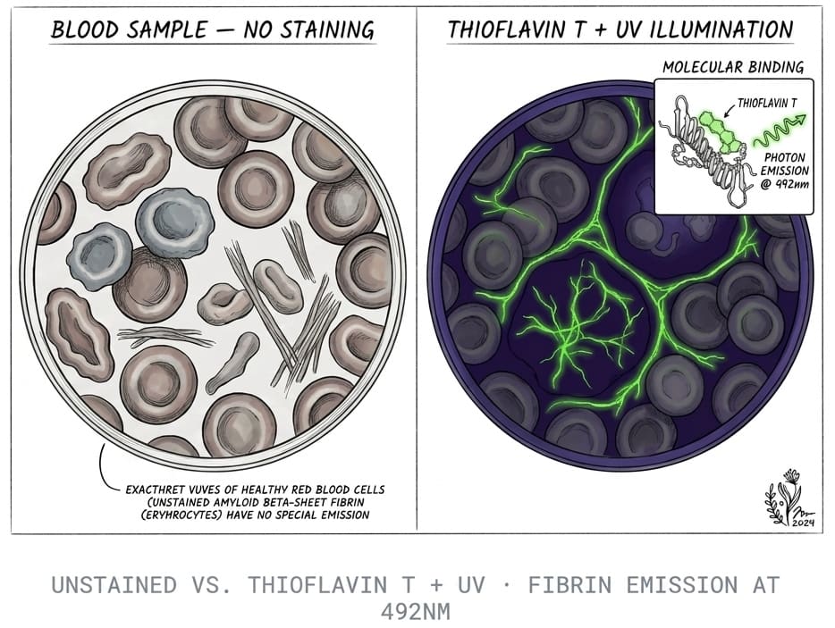
The only way to test for microclots today is to look at a blood or plasma sample stained with Thioflavin T, a dye that glows when it binds amyloid, under a fluorescence microscope or an imaging flow cytometer. Standard tests such as D-dimer, PT/INR and fibrinogen don't image microclots. You can get a microclot test through some clinics, research studies or an at-home kit. None of them is a validated diagnosis yet.
Key takeaways
- 1Microclot tests stain blood or plasma with Thioflavin T and image it, either by fluorescence microscopy or imaging flow cytometry.
- 2D-dimer, PT/INR, aPTT and fibrinogen measure other parts of clotting and can't confirm or rule out microclots.
- 3You can get tested through a clinic, a research study or an at-home finger-prick kit, and each uses a different sample type.
- 4There's no agreed cutoff, so a microclot result is observational data for a clinician to read alongside your symptoms and routine labs.
If you've read about microclots and want to know whether you have them, the first surprise is that your usual blood panel can't tell you. This guide walks through how microclots are actually tested for, what each option involves, and how to read a result without over- or under-reading it.
Why routine blood tests miss microclots
Routine clotting tests were built for other jobs. They tell a doctor how quickly your blood clots, how much clotting protein you have, or whether a large clot is breaking down somewhere. None of them takes a picture of what's floating in your plasma.
Fibrin amyloid microclots are a different kind of object. The research by Resia Pretorius, Douglas Kell and colleagues describes them as small fibrin particles that have folded into an amyloid-like shape and resist the body's clot-dissolving enzymes. Because they're particles, the way to find them is to look for them. If you're new to the topic, start with what microclots are and come back here.
| Test | What it measures | Can it detect microclots? |
|---|---|---|
| D-dimer | Fibrin fragments released as clots dissolve | No. See D-dimer vs microclot test |
| PT/INR and aPTT | How fast plasma clots | No |
| Fibrinogen | How much clotting protein is in plasma | No |
| Platelet count | Number of platelets | No, though platelet shape changes show up on microclot imaging |
| Thioflavin T microscopy | Fluorescent amyloid deposits in plasma or blood | Yes, this is the research method |
| Imaging flow cytometry with ThT | Count and size of fluorescent particles | Yes, a newer research method |
How do you test for microclots?
Every microclot test in use today rests on the same idea. Thioflavin T (ThT) is a dye that barely fluoresces in solution but lights up when it binds the stacked beta-sheets of amyloid fibrils. Add it to a blood sample, shine the right light on it, and amyloid deposits glow against a dark background.
What changes between tests is the sample and the way the glowing particles are counted.
Fluorescence microscopy
This is the original method. Platelet-poor plasma is stained with ThT and imaged under a fluorescence microscope. A reader then grades how many deposits there are, how big they are and how they're spread out. The Pretorius group used a grading system for microclots and platelet pathology and reported significant microclots in all 80 Long COVID samples it analyzed in that 2022 study.
The weak point is the reader. Grading by eye depends on which fields get chosen and who's looking. Recorded images or video help because a second person can check the same fields.
Imaging flow cytometry
To make counting more objective, the same group tested imaging flow cytometry with ThT. The sample flows past a camera one particle at a time, so the instrument counts and sizes every fluorescent particle in the sample. The authors wrote that their earlier microscopy lacked statistical strength, objectivity and throughput, which is a candid admission worth knowing about. These instruments are expensive, so the method is mostly found in research labs.
Automated image analysis
A 2024 preprint described software that measures fibrinaloid microclots automatically from microscope images. It's not peer reviewed yet, but it points to where testing is going: less judgment, more counting.
Where can you get a microclot test?
There are three realistic routes. Which one suits you depends on whether you want a clinical consultation, are happy to contribute to research, or mainly want data you can bring to your own doctor.
| Route | Sample | What you get | Good for |
|---|---|---|---|
| Long COVID or integrative clinic | Usually a venous blood draw | Imaging plus a consultation, often with other labs | People who want a clinician involved from the start |
| Research study | Venous blood draw | Usually no individual result | People who want to contribute data |
| At-home kit | Finger-prick dried blood sample | A report you can share | People who want data without a clinic visit |
Our directory of microclot testing labs and treatment centers lists clinic options by region and the questions to ask each one.
Testing at home
Our at-home amyloid test is the kit route. You collect a finger-prick dried blood sample and mail it to our partner laboratory in Mexico City. The lab stains it with Thioflavin T, the same staining principle used in the published microclot research, and records 6K 12-bit RAW video microscopy at 20× magnification under UV illumination. Footage is reviewed with AI-assisted fibril detection plus trained human oversight.
You get a Videographic Microscopy Report in a secure portal within 1 day of the lab receiving your sample, typically 5 to 7 business days from kit delivery including shipping. It costs $160 USD (3,200 MXN). The full method is in our guide to the amyloid fibrin test.
It's not a diagnostic test or a medical device. It doesn't measure spike protein, D-dimer or any standard clotting value, and a dried finger-prick sample isn't the same material as the platelet-poor plasma used in most published studies.
Next step
Ready to see your own sample?
Our at-home amyloid fibrin test uses Thioflavin T fluorescence microscopy and gives you a Videographic Microscopy Report to share with your doctor.
See the at-home testWhat should you ask before booking any microclot test?
The word "microclot test" covers quite different services, so compare them on the same points:
- Sample type. Venous plasma, whole blood and dried finger-prick samples aren't interchangeable, and results across them aren't directly comparable.
- Method. Is it ThT fluorescence microscopy, imaging flow cytometry, or something else? At what magnification?
- Who reads it. Manual grading, software counting and human review each have trade-offs. Ask whether you'll see the images or only a score.
- What the report claims. Walk away from any service that calls a microclot result a diagnosis, quotes an accuracy figure with no published study behind it, or ties the result to a treatment it sells.
- Whether your doctor can use it. A report only helps if a clinician can see what was done.
Before your test
Follow the collection instructions you're given and note the date, any recent infection, and your medicines and supplements, including enzymes like nattokinase. Don't stop prescribed anticoagulants or antiplatelet drugs to get a "cleaner" result. That's a decision for the doctor who prescribed them.
How to read a microclot test result
A report describes what was seen: whether ThT-positive deposits were present, roughly how many, how large, and sometimes how platelets looked. That's useful, but it has hard limits.
- There's no agreed normal range. Different labs grade differently, and no cutoff has been validated against clinical outcomes.
- It doesn't say what caused the deposits. Infection, inflammation, diabetes and other conditions have all been linked to microclots in the research.
- It doesn't diagnose a condition or prove that your symptoms come from microclots.
- The field is contested. Haematologists led by Beverley Hunt published a critique of the microclot hypothesis in 2024, and Kell, Khan and Pretorius replied defending the evidence. Read both if you want the full picture.
A positive result is a reason for a conversation with a clinician, not a treatment plan. If you're reading about options, our review of nattokinase and lumbrokinase for microclots sets out what the evidence does and doesn't support, and our doctor directory lists clinicians familiar with this research.
When not to use a microclot test
A microclot test isn't for emergencies. Sudden leg swelling, chest pain, sudden breathlessness, or stroke symptoms such as facial drooping or slurred speech need emergency care and standard tests like D-dimer and imaging. Don't wait for a kit result.
It's also not a test for Alzheimer's disease or systemic amyloidosis. Those use different methods, which we compare in types of amyloid tests.
Where to go next
If you have Long COVID and want the wider context, microclots and Long COVID explains how the research ties microclots to symptoms. If you want the chemistry behind the dye, the amyloid fibril test pillar compares Thioflavin T with Congo red and electron microscopy.
Frequently asked questions
Is there a blood test for microclots?
Yes, but not a standard one. Microclot tests use a blood or plasma sample stained with Thioflavin T and imaged under a fluorescence microscope or an imaging flow cytometer. They're offered by some clinics and by at-home kits, and they're used in research. None has regulatory approval as a diagnostic test, and there's no agreed reference range, so results are read as observational data.
Can my doctor order a microclot test?
Most hospital and commercial labs don't run microclot imaging, so it usually isn't on a standard order form. Some Long COVID and integrative clinics offer it directly. Your doctor can still review a report you bring from a clinic or a kit, and a report that includes images or video makes that conversation easier.
Will a D-dimer test show microclots?
No. D-dimer measures fragments released when clots break down and is mainly used to help rule out deep vein thrombosis and pulmonary embolism. The research describes fibrin amyloid microclots as resisting breakdown, so D-dimer answers a different question. A normal D-dimer doesn't rule microclots out, and a raised one doesn't confirm them.
How much does a microclot test cost?
Prices vary a lot between clinics and depend on what's bundled with the test, such as a consultation or other bloodwork. Our at-home amyloid fibrin test costs $160 USD (3,200 MXN) and includes a Videographic Microscopy Report you can share with any healthcare provider. Ask any provider what sample they use and what the report contains before comparing prices.
How long does a microclot test take?
Imaging itself is quick. The wait is mostly collection and shipping. With our kit, the report is ready within 1 day of the lab receiving your sample, and kit delivery to report typically takes 5 to 7 business days including shipping. Clinic turnaround depends on whether they image samples on site or send them out.
Sources
- A central role for amyloid fibrin microclots in long COVID/PASC: origins and therapeutic implications. Biochemical Journal, 2022
- Prevalence of symptoms, comorbidities, fibrin amyloid microclots and platelet pathology in individuals with Long COVID/PASC. Cardiovascular Diabetology, 2022
- Accelerating discovery: A novel flow cytometric method for detecting fibrin(ogen) amyloid microclots using long COVID as a model. Heliyon, 2023
- Automated microscopic measurement of fibrinaloid microclots and their degradation by nattokinase, the main natto protease. bioRxiv (preprint), 2024
- Molecular mechanism of Thioflavin-T binding to amyloid fibrils. Biochimica et Biophysica Acta, 2010
- D-dimer Test. MedlinePlus, U.S. National Library of Medicine, 2024
- Challenging the current hypothesis that thrombosis is responsible for the post-COVID-19 condition. Research and Practice in Thrombosis and Haemostasis, 2024
- Fibrinaloid microclots in long COVID: assessing the actual evidence properly. Research and Practice in Thrombosis and Haemostasis, 2024
At-Home Amyloid Test
Ready to see your own sample?
Our at-home amyloid fibrin test uses Thioflavin T fluorescence microscopy and gives you a Videographic Microscopy Report to share with your doctor.
Next steps
- What Are Microclots? A Plain-Language Guide to Amyloid MicroclotsWhat microclots are, how fibrin folds into amyloid microclots that resist breakdown, the symptoms researchers link to them, and how they're tested.
- Amyloid Fibrin Test: How Fibrin Amyloid Microclots Are DetectedWhat an amyloid fibrin test is, how Thioflavin T microscopy spots fibrin amyloid microclots, how it differs from D-dimer, and what it can't tell you.
- Amyloid Fibril Test: Methods Used to Detect Amyloid FibrilsHow an amyloid fibril test works: Thioflavin T, Congo red, electron microscopy, X-ray diffraction and seeding assays compared, with which are clinical.

Tim Ottowitz
Founder & Research Lead
Tim Ottowitz founded Amyloid Test and writes its Science Hub, turning fibrin amyloid and spike protein research into plain-language guides that stay close to the published evidence.
Keep reading
Amyloid Microclots: What the Research Shows So Far
What studies show about amyloid microclots in Long COVID: how fibrin forms them, why they resist breakdown, the key findings and the open questions.
D-Dimer vs Microclot Test: Why a Normal D-Dimer Can Miss Microclots
D-dimer vs microclot test compared: what each measures, why D-dimer can read normal in Long COVID, and how Thioflavin T microscopy finds fibrin amyloid.

Microclot Testing Labs and Treatment Centers: How to Choose
Compare microclot testing labs, research programs and apheresis or enzyme treatment options, with the questions to ask before you pay for any of them.