Best at Home Microclot Test Kits in 2026: An Evidence-Based Guide
English Narration
At-Home Blood Spot • 6K Video Microscopy • $160
Short answer
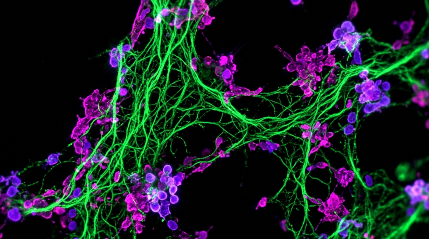
There is no FDA-cleared diagnostic test kit for microclots. The primary at-home option is a mail-in finger-prick collection kit using Thioflavin T fluorescence microscopy on stabilized dried blood. Hospital labs do not offer microclot imaging, and European apheresis clinics require in-person travel costing thousands of dollars.
Key takeaways
- 1At-home microclot testing relies on Thioflavin T fluorescence microscopy performed on mail-in dried blood samples.
- 2Routine hospital labs such as Quest or Labcorp run D-dimer and standard coagulation panels, but they do not image microclots.
- 3Laboratory analysis of dried-blood slides takes 24 to 48 hours, while full door-to-door transit takes 2 to 3 weeks via international courier.
- 4Evaluating a testing provider requires checking for negative controls, reagent blank slides, and access to raw microscopy video.
When persistent post-viral fatigue, brain fog, and breathlessness continue despite normal blood tests, many patients look into testing for anomalous blood clotting. Research by Resia Pretorius and Douglas Kell demonstrated that individuals with post-viral illness frequently harbor persistent, amyloid-like fibrin deposits known as microclots.
However, finding a reliable way to check for these structures is challenging. Standard commercial laboratories do not offer microclot assays, and specialized clinics in Europe require international travel. For many individuals, mail-in at-home collection kits provide an accessible alternative.
This guide reviews the current options for checking microclot burden, compares mail-in kits with clinical alternatives, and outlines the scientific criteria to check before ordering.
The Testing Landscape: How Options Compare
Patients seeking to evaluate microclot presence generally encounter three distinct pathways:
| Testing Option | Method | Specimen Type | Typical Cost | Total Turnaround |
|---|---|---|---|---|
| Mail-In At-Home Kit | ThT Fluorescence Microscopy (6K Video) | Dried blood on optical slide | $160 USD | 2 to 3 weeks (door-to-door) |
| European Apheresis Clinic | Fluorescence Microscopy / Flow Cytometry | Fresh venous platelet-poor plasma | $750 to $2,500+ | 1 to 2 days on site (requires travel) |
| Hospital Laboratory Panel | D-dimer, Fibrinogen, PT/INR | Liquid venous blood | $50 to $300 | 1 to 3 days (does not detect microclots) |
Understanding the differences between these approaches helps avoid misplaced expectations.
1. Mail-In At-Home Testing Kits
The primary commercial at-home option available is the amyloid fibrin test mail-in kit from Amyloid Test.
- How it works: You receive a kit at home containing sterile lancets, collection cards, and medical-grade glass slides. After collecting a small finger-prick blood drop, the sample dries on the slide, stabilizing the cellular architecture. You return the kit in a prepaid mailer.
- Laboratory processing: At the partner laboratory, technicians apply Thioflavin T (ThT), an optical fluorophore that binds selectively to beta-sheet protein aggregates. A specialized microscope captures high-resolution 6K video as the focal plane moves through the sample.
- Reporting: You receive a digital Videographic Microscopy Report containing raw video recordings and image documentation showing whether fluorescent amyloid microclots are present.
- Realistic timing: Laboratory imaging takes 24 to 48 hours once the specimen arrives. However, because kits travel via international courier and clear customs, the overall process from ordering to receiving results spans 2 to 3 weeks.
- Key advantage: It provides accessible, verifiable visual data without the necessity of traveling overseas.
2. Specialized In-Person Clinics
Several private clinics in Germany, Switzerland, and Cyprus evaluate microclots in person, often alongside treatments such as H.E.L.P. apheresis or triple therapy.
- How it works: Clinics draw fresh venous blood into citrated tubes, centrifuge it immediately to isolate platelet-poor plasma (PPP), and stain it on site with ThT.
- Pros: They use fresh liquid plasma, which matches the protocols used in early research papers.
- Cons: High total expense. In addition to clinic fees ($750 to $2,500), patients must pay for international flights and lodging while traveling with chronic fatigue.
3. Hospital and Commercial Lab Panels
A common point of confusion is whether a primary care doctor can order a microclot screen through Quest, Labcorp, or hospital laboratories.
Standard blood tests measure soluble clotting factors:
- D-dimer: Measures soluble fibrin fragments produced when normal clots degrade. As explained in our guide on D-dimer vs microclot testing, amyloid microclots resist natural fibrinolysis, meaning D-dimer is frequently normal even when microclot burden is elevated.
- Fibrinogen and PT/INR: Measure clotting factor levels and coagulation speed, not physical microclot particles.
Standard hospital networks do not maintain fluorescence microscopes calibrated for ThT imaging. For details on how methods differ, read how to test for microclots.
5 Criteria to Evaluate Before Ordering an At-Home Test
Because at-home microclot screening is an emerging observational service, quality varies significantly. Check these five technical factors before ordering:
1. Fluorophore Specificity (Thioflavin T)
Make sure the service specifically utilizes Thioflavin T (ThT). ThT is the reference standard in biophysical chemistry for detecting cross-beta sheet amyloid structures. When unbound in solution, ThT has low fluorescence; upon binding to amyloid cavities within misfolded fibrin, its fluorescence intensity increases substantially at 492nm.
2. Slide Fixation vs Liquid Transit
Sample stability during transit is critical. Liquid blood in unpreserved tubes degrades rapidly at ambient temperatures, causing red blood cells to lyse and potentially triggering artifactual clotting during shipping. Medical-grade dried smears on glass slides immobilize structures, ensuring that what the laboratory images reflects the initial sample rather than transit artifacts.
3. Negative Controls and Blank Slides
Any fluorescent dye can precipitate or bind nonspecifically if not carefully controlled. A rigorous laboratory runs verified negative control slides (healthy plasma) and reagent blank slides (dye without blood) in every batch to confirm background darkness before evaluating patient specimens.
4. Video Microscopy vs Static Stills
Static photos can be misleading, as dust particles or fiber fragments can mimic clots under single-frame illumination. Continuous 6K video microscopy tracks depth of field across the entire sample area, distinguishing true three-dimensional fibrin aggregates from surface artifacts.
5. Transparent Turnaround Expectations
Be wary of services claiming instantaneous turnaround times. While the physical staining and microscopy analysis takes 24 to 48 hours in the laboratory, safe transit across international borders takes time. A transparent provider explains that door-to-door delivery typically takes 2 to 3 weeks.
Understanding What Your Results Mean
A microclot screening report provides observational laboratory findings. It is not an FDA-cleared diagnostic test and does not constitute a clinical diagnosis of Long COVID or ME/CFS.
- If microclots are identified: The video footage provides visual documentation of structural fibrin aggregation. This data can be shared with a doctor familiar with microvascular research to discuss whether supportive therapies, such as nattokinase and lumbrokinase or anticoagulant protocols, are appropriate.
- If microclots are minimal or absent: It suggests anomalous fibrin aggregation may not be the primary driver of current symptoms, helping direct clinical investigations toward other mechanisms, such as mitochondrial dysfunction or viral persistence.
To explore how these findings fit into broader post-viral illness, review our detailed guide on what are microclots or consult our doctor directory to find clinicians experienced in managing post-viral coagulation issues.
Frequently asked questions
What is the best at home test kit for amyloid fibrin microclots in 2026?
The primary at-home testing option is the Amyloid Test finger-prick kit. It uses Thioflavin T (ThT) fluorescence microscopy on dried blood to image anomalous beta-sheet fibrin structures for $160 USD, featuring 6K video microscopy and laboratory analysis within 24 to 48 hours of specimen arrival.
How long does a mail-in microclot test take from order to report?
Laboratory analysis takes 24 to 48 hours once the specimen reaches the lab. However, because collection kits and blood samples travel via international couriers across customs, the full door-to-door cycle usually takes 2 to 3 weeks.
Can standard hospital laboratories test for amyloid microclots?
No. Standard hospital and commercial laboratories run soluble protein assays like D-dimer, PT/INR, and fibrinogen. They do not maintain calibrated fluorescence microscopes or ThT staining protocols to detect insoluble amyloid microclots.
What should I look for in a quality at-home microclot test?
Look for standardized Thioflavin T excitation and emission optics, verified negative batch controls to rule out dye precipitation, temperature-stable glass collection slides, and full access to raw video footage rather than a subjective numerical score.
Sources
- A central role for amyloid fibrin microclots in long COVID/PASC: origins and therapeutic implications. Biochemical Journal, 2022
- Prevalence of symptoms, comorbidities, fibrin amyloid microclots and platelet pathology in individuals with Long COVID/PASC. Cardiovascular Diabetology, 2022
- Molecular mechanism of Thioflavin-T binding to amyloid fibrils. Biochimica et Biophysica Acta, 2010
- Automated microscopic measurement of fibrinaloid microclots and their degradation by nattokinase. bioRxiv (preprint), 2024
At-Home Amyloid Test
Get Your Amyloid Test Kit Today
Simple at-home collection. Results in days.
Next steps
- What Are Microclots? A Plain-Language Guide to Amyloid MicroclotsWhat microclots are, how fibrin folds into amyloid microclots that resist breakdown, the symptoms researchers link to them, and how they're tested.
- How to Test for Microclots: Methods, Options and What Results MeanHow to test for microclots: the methods researchers use, why D-dimer can't detect them, where to get a microclot test and how to read the result.
- Amyloid Fibrin Test: How Fibrin Amyloid Microclots Are DetectedWhat an amyloid fibrin test is, how Thioflavin T microscopy spots fibrin amyloid microclots, how it differs from D-dimer, and what it can't tell you.

Tim Ottowitz
Founder & Research Lead
Tim Ottowitz founded Amyloid Test and writes its Science Hub, turning fibrin amyloid and spike protein research into plain-language guides that stay close to the published evidence.
Keep reading
Amyloid Microclots: What the Research Shows So Far
What studies show about amyloid microclots in Long COVID: how fibrin forms them, why they resist breakdown, the key findings and the open questions.
Anomalous Amyloid Microclots: Phenotypes and Phosphatidylserine
Explore the biophysics of anomalous amyloid microclots. Discover the 4 structural phenotypes, phosphatidylserine membrane flipping, and ThT microscopy.
Microclots Symptoms: Capillary Occlusion, Fatigue, and Dyspnea
Understand key microclots symptoms in blood and tissue. Learn why capillary hypoperfusion causes fatigue, brain fog, and breathlessness with normal labs.