Finger-Prick Amyloid Test: How Two Blood Drops Detect Fibrils
Short answer
Two drops of capillary blood (50 to 100 microliters) can detect circulating vascular amyloid fibrils because peripheral capillary beds mechanically concentrate large, fibrinolysis-resistant aggregates, while Thioflavin T fluorescence amplifies optical signal up to 1,000-fold. Key takeaway: Capillary samples reflect circulating microvascular microclots, but they cannot diagnose organ-deposited systemic amyloidosis or replace tissue biopsies.
Key takeaways
- 1Capillary beds in the fingertip act as mechanical filters (5 to 10 micrometers) that trap circulating fibrin amyloid microclots measuring 2 to 200 micrometers.
- 2Because blood completes a systemic circuit every 60 seconds, peripheral blood reflects body-wide exposure to inflammatory and amyloidogenic triggers.
- 3Thioflavin T fluorescence jumps 50- to 1,000-fold upon binding cross-beta sheet grooves, making picogram-level fibril deposits visible on a microscope slide.
- 4Finger-prick testing distinguishes circulating vascular microclots from tissue-locked systemic amyloidosis (AL/ATTR), which still requires organ biopsy.
- 5Capillary sampling introduces technical artifacts like tissue factor activation and dehydration clumping that demand specialized laboratory protocols.
If you have looked into testing for persistent vascular inflammation, you have likely encountered a surprising claim: two drops of blood from a routine finger prick can show whether amyloid fibrils are circulating in your body. At first glance, relying on a 50 to 100 microliter drop seems inadequate when evaluating a complex, body-wide illness. In this guide, you will learn the biophysical reasons why peripheral capillary blood concentrates circulating microclots, how modern optical stains detect them, and why finger-prick tests cannot replace clinical tissue biopsies for systemic organ amyloidosis.
Whether a finger-prick blood sample provides credible evidence depends on the clinical context. When applied to circulating fibrin amyloid microclots, the physics of capillary filtration and fluorophore amplification make two drops an informative window into your vascular health.
The Biophysical Mechanism: Why Capillary Blood Traps Circulating Fibrils
When an assay checks for fibrinogen that has misfolded into beta-sheet-rich, fibrinolysis-resistant microclots, two drops of capillary blood provide a surprisingly high-density specimen. Three distinct physiological mechanisms explain why this works.
1. Capillary Rheology and Mechanical Trapping
The internal diameter of arterioles and capillaries in human fingertips measures roughly 5 to 10 micrometers (µm). Red blood cells, which are about 7 to 8 µm in diameter, deform into bullet shapes to squeeze through these tight lumens in single file.
In contrast, circulating fibrin amyloid aggregates typically span 2 to 200 µm across. Unlike standard physiologic blood clots, which plasmin dissolves within hours, these anomalous deposits contain dense cross-beta sheet architecture that resists enzymatic degradation. Because these rigid structures cannot fold or compress, they slow down, collide with vessel walls, and mechanically lodge within peripheral microvascular networks. The capillary beds of the fingertip act as a natural physical filter, concentrating circulating insoluble aggregates that remain diluted in large high-flow veins.
2. Rapid Systemic Mixing of Soluble Precursors
The entire adult blood volume (roughly 5 liters) completes a full circulatory loop through the heart and vasculature approximately once every 60 seconds. Because blood circulates so rapidly, any systemic inflammatory trigger in the circulation contacts the entire soluble fibrinogen pool almost continuously.
When circulating inflammatory mediators, such as bacterial lipopolysaccharide (LPS), acute-phase proteins like Serum Amyloid A, or viral proteins, trigger conformational misfolding, they do so throughout the entire circulating blood volume. Soluble fibrinogen molecules across the systemic circulation undergo abnormal polymerization into anomalous aggregates simultaneously. A peripheral capillary blood sample taken from a finger prick accurately captures the biochemical environment of the broader vascular pool.
3. Fluorophore Amplification via Thioflavin T
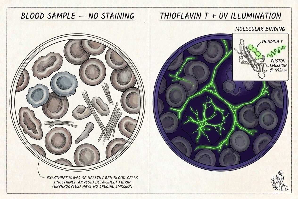
In fluorescence microscopy assays, the blood sample is stained with Thioflavin T (ThT), a benzothiazole fluorophore dye. The molecular properties of ThT create massive signal amplification:
- In free aqueous solution: The benzylamine and benzothiazole aromatic rings of the ThT molecule rotate freely around their shared carbon-carbon bond. When excited by light, this rotation dissipates energy without emitting photons, resulting in virtually zero background fluorescence.
- Upon binding amyloid fibrils: ThT molecules slip into the narrow channel grooves running parallel to the long axis of cross-beta sheets. This steric confinement locks the molecule into a rigid planar configuration, blocking non-radiative rotational decay.
- Fluorescence emission jump: When illuminated by blue excitation light (~440 to 450 nm), the locked dye emits intense green-yellow light (~480 to 490 nm). Its fluorescence quantum yield jumps between 50-fold and 1,000-fold compared to unbound solution.
Because of this dramatic signal-to-noise multiplication, a laboratory camera does not need large volumes of protein to detect abnormal structures. A standard 20x objective lens scanning a single drop of stained capillary blood can detect tiny picogram quantities of anomalous protein aggregates as bright glowing structures.
Three Distinct Testing Contexts for Amyloid Exposure
Much of the confusion around finger-prick testing comes from the term "amyloid." In clinical medicine, amyloid refers to any protein that misfolds into insoluble cross-beta sheet fibrils. However, the location and identity of those fibrils dictate whether a two-drop finger-prick sample provides meaningful data.
The table below contrasts how a capillary blood sample performs across three separate testing categories:
| Testing Context | Target Analyte | Detection Method | Is 2 Drops Enough Evidence? | Clinical Role |
|---|---|---|---|---|
| 1. Fibrin Amyloid Microclots | Misfolded fibrin cross-beta aggregates in blood | Thioflavin T (ThT) fluorescence microscopy | Yes, for circulating vascular load. Capillary beds concentrate these micro-aggregates, making them visible under 20x optical magnification. | Research-grade marker for persistent vascular inflammation in chronic conditions. |
| 2. Alzheimer's Neuro-Biomarkers | Soluble Aβ42/Aβ40 peptides and phosphorylated tau (p-tau217) | Ultrasensitive digital immunoassay (Simoa) or mass spectrometry | Yes, as a surrogate risk biomarker. Digital ELISA detects femtogram-per-milliliter concentrations in dried blood spots. | Remote screening tool that correlates with brain amyloid PET scans and cognitive decline. |
| 3. Classic Systemic Amyloidosis | Insoluble amyloid light-chain (AL) or transthyretin (ATTR) in organs | Congo red staining under polarized light on tissue biopsy | No. Organ-deposited fibrils remain trapped inside myocardium, renal glomeruli, or nerves, not circulating freely. | Diagnostic standard requiring heart, kidney, or abdominal fat pad tissue biopsy. |
Understanding these distinctions prevents misinterpreting your results. If you are comparing tests, explore our overview of types of amyloid tests and our deep dive into the amyloid fibrin test methodology.
Technical Limitations: Where Finger-Prick Sampling Breaks Down
While capillary blood is remarkably efficient for detecting circulating vascular microclots, finger-prick collection introduces technical variables that don't exist in standard hospital venipuncture. Clinicians and researchers evaluate four key limitations:
Capillary Trauma vs. Centrifuged Platelet-Poor Plasma
The peer-reviewed laboratory protocols established by Resia Pretorius, Douglas Kell, and colleagues were developed using venous blood collected in sodium citrate tubes and centrifuged into Platelet-Poor Plasma (PPP). Centrifugation removes cellular elements (red blood cells and platelets), isolating pure plasma for microscopy.
When you perform a finger prick, the lancet pierces skin capillaries and surrounding tissue, releasing tissue factor (thromboplastin). Tissue factor activates coagulation factor VII, triggering the extrinsic clotting pathway. Additionally, if a patient squeezes or "milks" the fingertip aggressively to produce blood, the mechanical pressure shears red cells and activates platelets. When blood drops dry on a glass slide or collection card, fluid evaporation creates Marangoni flow ("coffee-ring" effect), which can drag proteins into clumps. Specialized laboratories account for this by training patients on clean collection and using standardized morphological criteria to distinguish genuine amyloid microclots from collection artifacts.
Conformation Specificity vs. Protein Identity
Thioflavin T is conformation-specific, not protein-specific. It recognizes the spatial geometry of cross-beta sheet architecture, regardless of which parent protein assembled the structure.
A glowing green signal on a capillary blood smear confirms the presence of beta-sheet amyloid fibrils in the specimen. However, ThT alone cannot determine whether those fibrils originated from misfolded fibrinogen, Serum Amyloid A, broken cellular scaffolding, or foreign peptides. Confirming the exact protein composition requires secondary methods such as immunofluorescent antibody labeling or liquid chromatography tandem mass spectrometry.
Autofluorescence and Cellular Background
Whole capillary blood contains hemoglobin, porphyrins, flavins, and oxidized lipid membranes. Several of these biological compounds exhibit natural autofluorescence when excited by blue or ultraviolet light.
In whole dried blood smears, cellular debris can produce faint background illumination. Trained microscopists and automated computer-vision algorithms prevent false positives by applying specific thresholding: they evaluate particle size, boundary sharpness, and emission intensity to separate genuine ThT-bound microclots from background cellular autofluorescence.
Circulating Shedding vs. Tissue-Bound Pathology
Amyloid fibrils possess hydrophobic surfaces that make them naturally adherent. In many chronic vascular conditions, the most problematic aggregates do not circulate continuously. Instead, they adhere to the endothelial glycocalyx lining blood vessel walls or incorporate into microvascular beds throughout deep tissues.
A finger prick samples only what is actively floating in the peripheral bloodstream at the exact moment of collection. A clear sample means circulating fibril concentrations in capillary blood are low; it does not guarantee that microvascular walls or extravascular organs are entirely free of deposited material.
Where Our At-Home Amyloid Test Fits
To address the barrier of expensive, travel-heavy clinical evaluations, our at-home amyloid test brings research-grade microvascular analysis directly to patients.
Here is how our collection and analysis pipeline works:
- At-home dried blood collection: You collect a small capillary blood sample using a sterile lancet and mail it directly to our partner laboratory in Mexico City.
- Laboratory preparation and ThT staining: Technicians treat the sample with Thioflavin T to label any cross-beta sheet amyloid structures present.
- 6K 12-bit RAW video microscopy: The lab records high-resolution digital video across multiple slide fields at 20x optical magnification under ultraviolet illumination.
- AI-assisted detection with expert oversight: Trained analysts evaluate the footage using computer-vision models to quantify microclot surface area and structural density.
- Videographic Microscopy Report: Within one business day of the laboratory receiving your sample (typically 5 to 7 business days from ordering), you receive an objective video report in a secure client portal.
- Clear cost structure: The test costs $160 USD (3,200 MXN), avoiding the multi-thousand-dollar fees commonly charged by private specialty clinics.
What Our Test Does Not Do
To ensure safe and responsible interpretation, we maintain strict boundaries regarding what the Amyloid Test kit provides:
- It is not a diagnostic test or medical device: It provides observational laboratory data for research, informational, and educational tracking. It does not replace medical consultation with your physician.
- It does not measure active spike protein: Visualizing amyloid microclots evaluates downstream vascular consequences, but it does not quantify circulating spike antigen or antibody levels.
- It is not an Alzheimer's test: Our microscopy assesses microvascular fibrin aggregates, not cerebrospinal fluid peptides, plasma p-tau217, or brain amyloid PET plaques.
- It does not diagnose systemic organ amyloidosis: AL and ATTR amyloidosis require formal clinical workups, including organ biopsies, serum free light chain assays, and genetic testing.
- It does not replace standard coagulation panels: It does not measure traditional parameters like INR, PT, PTT, or D-dimer.
If you are evaluating clinic options, review our comparison of at-home microclot tests vs clinic testing and our analysis of flow clotometry vs microscopy.
Next Steps for Patients and Clinicians
Two blood drops from a fingertip cannot diagnose classic organ amyloidosis, but the physics of capillary filtration and fluorophore amplification make them a practical, sensitive method for observing circulating fibrin amyloid microclots.
If you are dealing with unexplained post-viral fatigue, exercise intolerance, or cognitive symptoms, sharing observational microscopy data with an open-minded healthcare provider can help guide your investigation. Start by reading our foundational guide on what microclots are and review the primary research in our amyloid fibrin test documentation.
Frequently asked questions
Can two drops of blood diagnose systemic amyloidosis like AL or ATTR?
No. Classic systemic amyloidosis involves insoluble fibril deposition directly inside solid organs like the heart, kidneys, and liver, or subcutaneous fat. A peripheral blood drop cannot measure organ-locked fibril burden or replace diagnostic tissue biopsies and Congo red histology.
Why do amyloid microclots collect in fingertip capillary blood?
Fingertip capillaries have narrow internal lumens of 5 to 10 micrometers. Circulating fibrin amyloid microclots typically span 2 to 200 micrometers and resist enzymatic breakdown by plasmin. This size mismatch causes rigid beta-sheet aggregates to lodge and concentrate in peripheral microvascular beds.
How does Thioflavin T make tiny amounts of amyloid visible?
In free solution, Thioflavin T molecules rotate freely and dissipate energy without glowing. When dye molecules bind into the parallel grooves of cross-beta sheet fibrils, their rotation locks. Under blue light excitation (440 to 450 nm), green emission fluorescence jumps 50- to 1,000-fold.
Does finger squeezing create false microclots on the slide?
Excessive squeezing shears skin cells and releases tissue factor (thromboplastin), which initiates rapid extrinsic coagulation. Furthermore, improper drying can cause phase-separation artifacts. Standardized protocols avoid aggressive milking, use clean lancet punctures, and control drying conditions to prevent non-specific clumping.
How does a finger-prick microclot test compare to an Alzheimer's blood test?
An amyloid microclot test uses fluorescence microscopy to view intact structural aggregates of misfolded fibrin. An Alzheimer's dried blood spot test uses ultrasensitive digital immunoassays (such as Simoa) or mass spectrometry to quantify soluble peptide ratios (Abeta42/40 and p-tau217) reflecting brain pathology.
Sources
- A central role for amyloid fibrin microclots in long COVID/PASC: origins and therapeutic implications. Biochemical Journal, 2022
- SARS-CoV-2 spike protein S1 induces fibrin(ogen) resistant to fibrinolysis. Biochemical Journal, 2021
- The Mechanism of Thioflavin-T Binding to Amyloid Fibrils. Journal of Molecular Biology, 2010
- Capillary blood dried blood spot testing for Alzheimer's disease biomarkers: The DROP-AD study. Nature Medicine, 2024
- Systemic Amyloidosis: Pathogenesis and Diagnostic Approaches. New England Journal of Medicine, 2003
- Prevalence of amyloid microclots in post-acute sequelae of COVID-19. Cardiovascular Diabetology, 2024
At-Home Amyloid Test
Check for circulating fibrin amyloid microclots
Order our $160 at-home collection kit, mailed to our partner lab in Mexico City for 6K RAW video fluorescence microscopy.
Next steps
- Amyloid Fibrin Test: How Fibrin Amyloid Microclots Are DetectedWhat an amyloid fibrin test is, how Thioflavin T microscopy spots fibrin amyloid microclots, how it differs from D-dimer, and what it can't tell you.
- Amyloid Fibril Test: Methods Used to Detect Amyloid FibrilsHow an amyloid fibril test works: Thioflavin T, Congo red, electron microscopy, X-ray diffraction and seeding assays compared, with which are clinical.
- Types of Amyloid Tests: Blood Tests, PET, CSF, Biopsy and MicroclotsCompare the types of amyloid tests: Alzheimer's blood tests, amyloid PET, spinal fluid, amyloidosis biopsy and fibrin amyloid microclot microscopy.

Tim Ottowitz
Founder & Research Lead
Tim Ottowitz founded Amyloid Test and writes its Science Hub, turning fibrin amyloid and spike protein research into plain-language guides that stay close to the published evidence.
Keep reading
Microclot Testing Access and Cost Index 2026
Compare at-home kits, private clinics, research studies and hospital testing: cost, sample, turnaround and what each route can actually show.
D-Dimer vs Microclot Test: Why a Normal D-Dimer Can Miss Microclots
D-dimer vs microclot test compared: what each measures, why D-dimer can read normal in Long COVID, and how Thioflavin T microscopy finds fibrin amyloid.
Amyloid Microclots Blood Test: Methods, Costs, and Home Kits
How to get an amyloid microclots blood test. Compare hospital research protocols with at-home ThT fluorescence microscopy kits, pricing, and turnaround.