D-Dimer vs Microclot Test: Why a Normal D-Dimer Can Miss Microclots
Short answer
D-dimer measures fragments released when the body breaks down a normal fibrin clot, so it's built to flag active clotting such as a DVT or pulmonary embolism. Fibrin amyloid microclots described in Long COVID research resist that breakdown, so they may not raise D-dimer at all. A microclot test instead stains plasma with Thioflavin T and looks for the clots directly under a fluorescence microscope.
Key takeaways
- 1D-dimer is a breakdown product, so it reflects clots that are being dissolved, not every clot that exists.
- 2Fibrin amyloid microclots resisted fibrinolysis in Long COVID plasma studies, which is one proposed reason D-dimer can stay normal.
- 3Thioflavin T fluorescence microscopy looks for the amyloid-rich clot material itself rather than its breakdown fragments.
- 4A normal D-dimer still matters: it's a useful tool for ruling out acute clots and should be read by a clinician.
- 5Microclot microscopy is a research-derived method, not a validated diagnostic standard, so results are observational data.
If your D-dimer came back normal but you still feel unwell after COVID-19, you're not alone, and the result isn't necessarily a contradiction. This guide compares D-dimer with microclot testing: what each one measures, why they can disagree, and what a normal result does and doesn't rule out.
What's the difference between a D-dimer and a microclot test?
They look at different things. D-dimer is an immunoassay that measures a fragment released when the enzyme plasmin breaks down cross-linked fibrin. A microclot test stains plasma with Thioflavin T (ThT) and looks for intact, amyloid-rich clot particles under a fluorescence microscope, the method described in the amyloid fibrin test guide.
| Feature | Standard D-dimer | Microclot test (ThT microscopy) |
|---|---|---|
| What it targets | Soluble fragments of broken-down fibrin | Intact fibrin amyloid particles that bind ThT |
| Method | Laboratory immunoassay | Fluorescence microscopy after ThT staining |
| What a high result suggests | Active clot formation and breakdown, such as DVT or PE | More amyloid-rich clot material in the sample |
| Main clinical use | Helping rule out acute venous clots | Research and observational data, not an accepted diagnostic |
| Reference range | Established lab cut-offs | No agreed clinical reference range yet |
| Sample | Venous blood draw | Plasma in research; some services use a finger-prick sample |
| Further reading | Types of amyloid tests | Fibrin amyloid microclots research |
Why can D-dimer be normal when microclots are present?
The short version: D-dimer counts what's being cleared, not what's stuck. If a clot resists breakdown, it won't release much D-dimer.
D-dimer measures clot breakdown, not clot presence
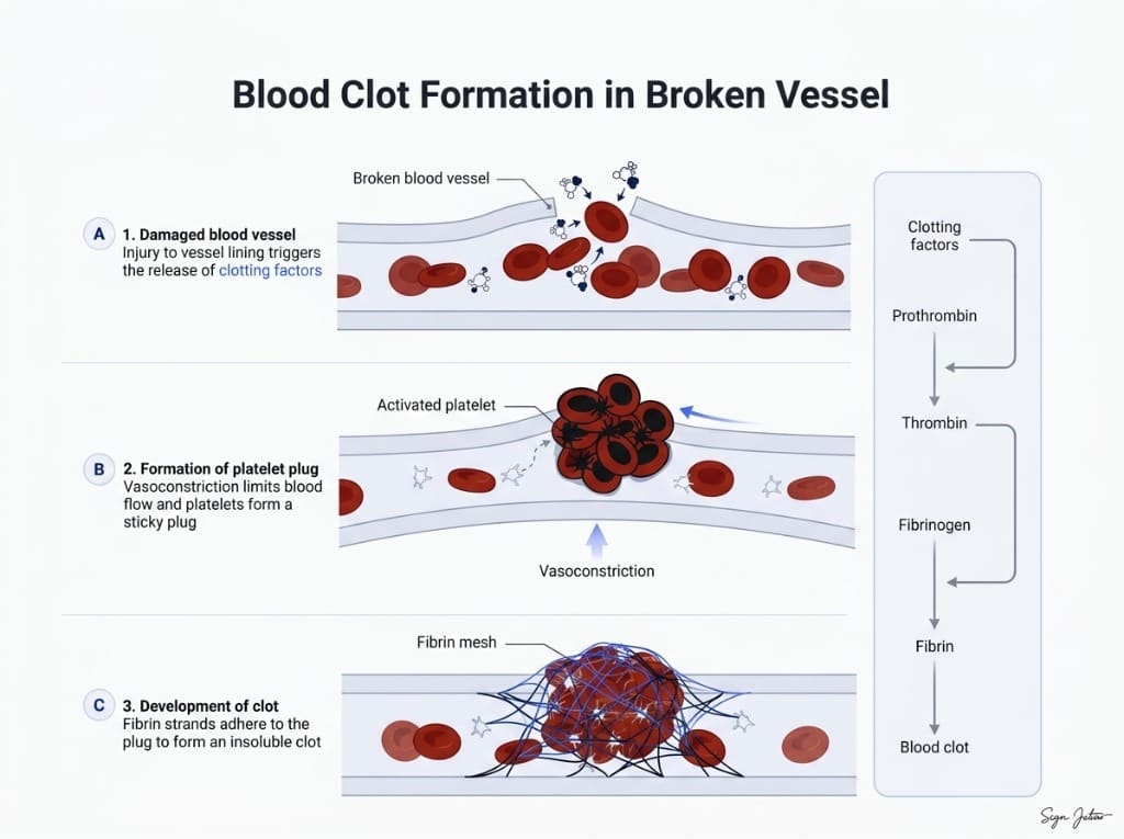
In a normal clot, thrombin turns fibrinogen into fibrin and Factor XIIIa cross-links it into a stable mesh. Tissue plasminogen activator then switches on plasmin, which cuts that mesh into soluble pieces. One of those pieces carries the D-dimer domain, and that's what the lab assay picks up.
So D-dimer rises when the body is actively forming and dissolving clots. That's why it's useful for ruling out a deep vein thrombosis or pulmonary embolism.
Fibrin amyloid microclots resist plasmin
Work led by Resia Pretorius and Douglas Kell found that plasma from people with Long COVID contained anomalous, amyloid-rich clot deposits that resisted fibrinolysis in the lab, even after a first round of trypsin digestion (Pretorius et al., 2021). Those deposits trapped inflammatory molecules, including raised levels of α2-antiplasmin, the body's main plasmin inhibitor.
Separate lab work showed that adding the spike protein S1 subunit to healthy plasma produced fibrin that was harder to break down (Grobbelaar et al., 2021), and test-tube work showed that fragments of spike can form amyloid fibrils themselves (Nyström and Hammarström, 2022). That mechanism is covered in more depth in our piece on spike protein amyloid fibrils and fibrinolysis.
If plasmin can't cut these clots efficiently, they won't shed much D-dimer. That's the leading explanation for a normal D-dimer alongside persistent symptoms, though it's a hypothesis from small observational and lab studies rather than a settled clinical fact (Kell et al., 2022).
What a normal D-dimer still tells you
A normal D-dimer is genuinely reassuring about one thing: an acute venous clot is less likely. It doesn't rule out microclots, and it doesn't prove them either. Some people do have raised D-dimer after COVID-19, and that result needs prompt medical follow-up.
Clinical next step
Request a clinical screening review
Share your case details and the ClearAmyloid team will review whether the protocol is a fit.
Start screeningHow Thioflavin T microscopy detects microclots
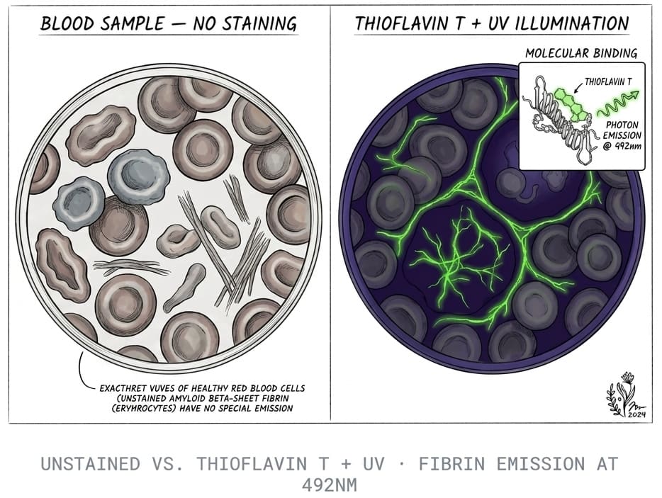
Thioflavin T is a fluorescent dye that's been used to stain amyloid since the late 1950s. In solution it barely glows, because its two ring structures spin freely and lose energy. When it slots into the grooves of beta-sheet-rich amyloid fibrils, that rotation stops and it fluoresces brightly under blue or UV light.
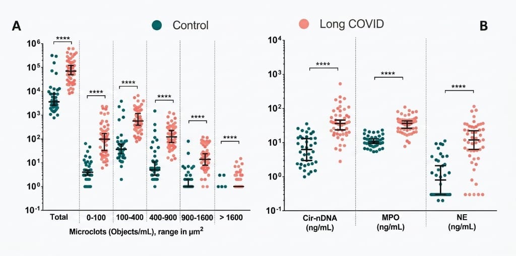
In the published studies, plasma from healthy controls showed few, small ThT-positive particles. Plasma from people with Long COVID showed more and larger fluorescent deposits (Pretorius et al., 2021). If you want the dye chemistry in detail, the amyloid fibril test guide walks through how ThT binding works and where it can mislead.
Researchers have since reported that these microclots are tangled up with neutrophil extracellular traps, which may help stabilize them. That link is explored in microclots and NETs in Long COVID.
Which symptoms lead people to ask about microclot testing?
People usually ask about microclot testing after standard bloods, including D-dimer, a full blood count and routine coagulation tests, have come back normal while symptoms continue. Common reasons include:
- Post-exertional malaise: crashes 12 to 72 hours after modest effort.
- Brain fog: trouble finding words, slower thinking or memory lapses.
- Breathlessness with normal oxygen readings.
- Cold hands and feet, tingling or skin colour changes.
- Persistent fatigue that rest doesn't fix.
None of these symptoms is specific to microclots. Researchers have proposed that poor capillary blood flow could contribute, but that link hasn't been proven in controlled trials. For a plain-language overview of the hypothesis, see amyloid microclots in Long COVID.
Where the Amyloid Test kit fits
Our at-home amyloid test uses the same ThT staining principle as the published microclot research. You collect a finger-prick dried blood sample and mail it to a partner laboratory in Mexico City. The lab records ThT fluorescence video microscopy and reviews it with AI-assisted fibril detection plus trained human oversight.
You get a Videographic Microscopy Report you can share with any healthcare provider. It isn't a diagnostic test, it doesn't measure D-dimer or any coagulation value, and it doesn't replace urgent assessment for a suspected DVT or PE.
Next steps after a normal D-dimer
Start by keeping the result. It's useful evidence that an acute clot was less likely at the time it was taken. If symptoms persist, a clinician who knows post-viral illness can help decide what else is worth checking.
If symptoms are wearing you down while you wait for answers, the Long COVID survival guide covers pacing and tracking. If you're weighing options, how to test for microclots compares the methods, and our guide to microclot testing labs and treatment centers compares where testing is available. For what's been studied once microclots are found, read the evidence on nattokinase for microclots. And if spike protein is part of your question, spike protein persistence testing explains why that needs a separate assay.
Frequently asked questions
Can you have microclots with a normal D-dimer?
Researchers studying Long COVID have reported fibrin amyloid microclots in plasma from people whose routine coagulation results didn't explain their symptoms. Because these clots resisted breakdown in lab studies, they may not release much D-dimer. That's a research finding, not a clinical rule, so a normal D-dimer doesn't prove microclots are present either. It simply means D-dimer and microclot microscopy answer different questions.
Is D-dimer ever elevated in Long COVID?
Yes, some people do show raised D-dimer after COVID-19, especially in the months after infection or after a severe acute illness. Others have normal results. A raised D-dimer needs prompt medical review because it can point to a deep vein thrombosis or pulmonary embolism, which are emergencies that a microclot test isn't designed to catch.
What blood test shows microclots?
No routine hospital blood test reports fibrin amyloid microclots. In the published research, Pretorius, Kell and colleagues stained platelet-poor plasma with Thioflavin T and imaged it with fluorescence microscopy. Some labs now offer the same staining principle outside research settings, but there's no agreed clinical reference range yet, so results are best treated as observational data for your clinician.
Should I ask my doctor for a D-dimer or a microclot test?
They're not substitutes. If you have sudden leg swelling, chest pain or breathlessness, ask for urgent assessment, which usually includes D-dimer and imaging. If acute clots are ruled out and symptoms persist, some patients choose a microclot test to add another data point. Bring any results to a clinician who can interpret them alongside your history.
Sources
- Persistent clotting protein pathology in Long COVID/Post-Acute Sequelae of COVID-19 (PASC) is accompanied by increased levels of antiplasmin. Cardiovascular Diabetology, 2021
- SARS-CoV-2 spike protein S1 induces fibrin(ogen) resistant to fibrinolysis: implications for microclot formation in COVID-19. Bioscience Reports, 2021
- A central role for amyloid fibrin microclots in long COVID/PASC: origins and therapeutic implications. Biochemical Journal, 2022
- Amyloidogenesis of SARS-CoV-2 Spike Protein. Journal of the American Chemical Society, 2022
At-Home Amyloid Test
Get Your Amyloid Test Kit Today
Simple at-home collection. Results in days.
Next steps
- Amyloid Fibrin Test: How Fibrin Amyloid Microclots Are DetectedWhat an amyloid fibrin test is, how Thioflavin T microscopy spots fibrin amyloid microclots, how it differs from D-dimer, and what it can't tell you.
- Amyloid Fibril Test: Methods Used to Detect Amyloid FibrilsHow an amyloid fibril test works: Thioflavin T, Congo red, electron microscopy, X-ray diffraction and seeding assays compared, with which are clinical.
- Types of Amyloid Tests: Blood Tests, PET, CSF, Biopsy and MicroclotsCompare the types of amyloid tests: Alzheimer's blood tests, amyloid PET, spinal fluid, amyloidosis biopsy and fibrin amyloid microclot microscopy.
Tim Ottowitz
Keep reading
Amyloid Microclots: What the Research Shows So Far
What studies show about amyloid microclots in Long COVID: how fibrin forms them, why they resist breakdown, the key findings and the open questions.
How to Test for Microclots: Methods, Options and What Results Mean
How to test for microclots: the methods researchers use, why D-dimer can't detect them, where to get a microclot test and how to read the result.
What Are Microclots? A Plain-Language Guide to Amyloid Microclots
What microclots are, how fibrin folds into amyloid microclots that resist breakdown, the symptoms researchers link to them, and how they're tested.