Amyloid Fibril Test: Methods Used to Detect Amyloid Fibrils
Short answer
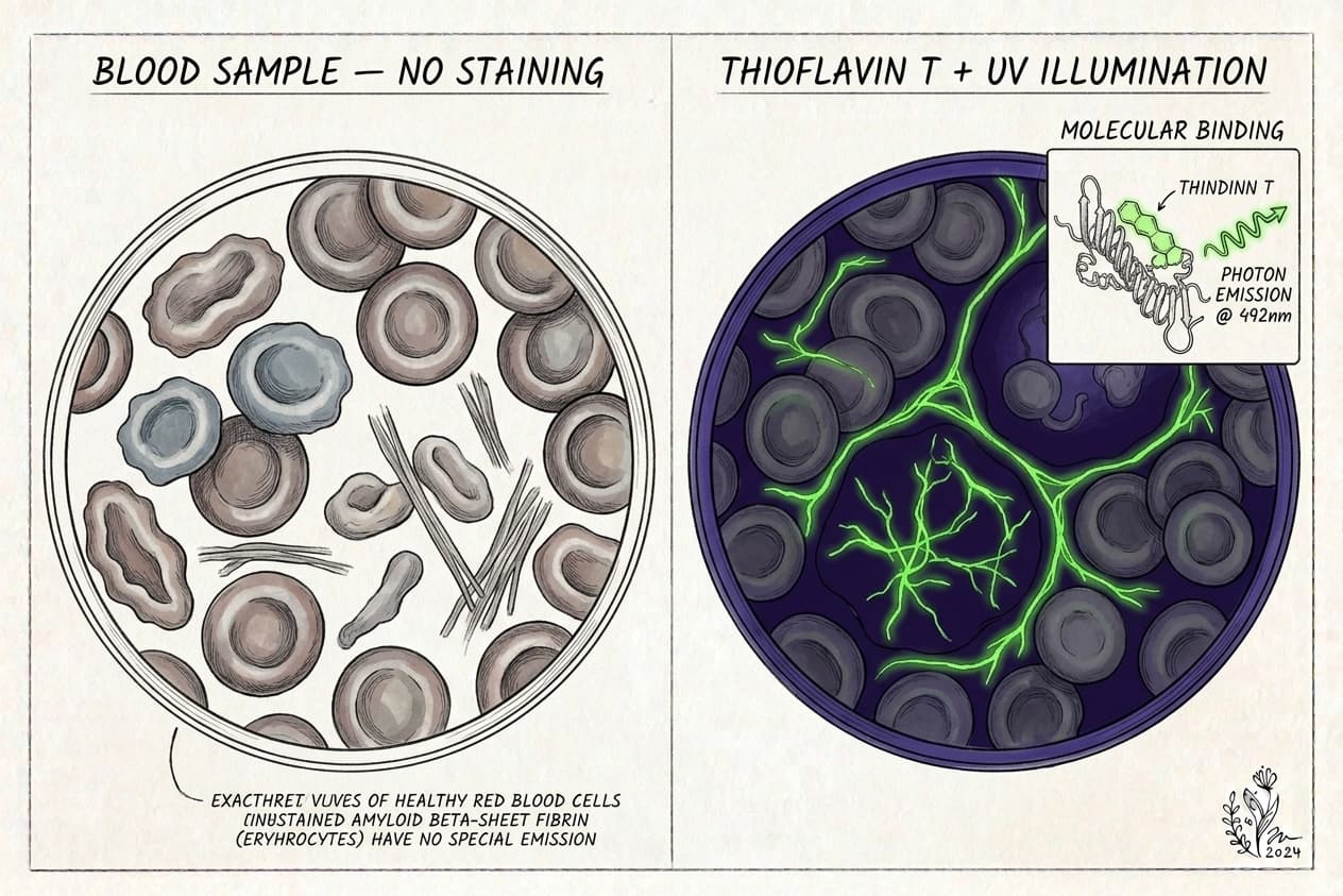
There isn't one amyloid fibril test. Scientists combine methods: Thioflavin T fluorescence to flag beta-sheet fibrils, Congo red birefringence in tissue, electron microscopy to see fibril shape, X-ray diffraction or cryo-EM to confirm the cross-beta structure, seeding assays like RT-QuIC to amplify tiny amounts, and antibodies for specific forms. Only a few, such as Congo red tissue staining and RT-QuIC, are routine clinical tools.
Key takeaways
- 1Amyloid is a shared protein shape (stacked beta-sheets in a cross-beta pattern), so tests look for that structure rather than one specific protein.
- 2Thioflavin T fluorescence is the most widely used screening method, but a ThT signal alone doesn't prove a deposit is amyloid.
- 3Congo red staining of tissue biopsies is the standard clinical stain for systemic amyloidosis, and it's usually paired with protein typing.
- 4Electron microscopy, X-ray diffraction and cryo-EM confirm fibril structure but are research tools, not routine blood tests.
- 5The spike protein amyloid studies and the fibrin amyloid microclot studies both rely on ThT, combined with other methods to build confidence.
If you've read about amyloid in Alzheimer's, in Long COVID microclots or in spike protein research, you've seen the same few lab techniques come up again and again. This guide explains what each amyloid fibril test actually detects, which ones you'd meet in a hospital and which stay in research labs, and how Thioflavin T gets applied to blood.
What is an amyloid fibril?
An amyloid fibril is a protein that has refolded into long, thin threads built from stacked beta-sheets. The strands run across the fibril's long axis, a pattern called cross-beta. In a 1997 synchrotron X-ray study, six different amyloid fibrils taken from tissue and two synthetic ones all gave similar diffraction patterns, which is strong evidence that amyloid is a shared shape rather than a single protein.
That's why "amyloid fibril test" is really a family of methods. Each one targets a different property of the shape: how it binds dyes, how it looks under a microscope, how it scatters X-rays or whether it can seed more of itself. Many proteins can take this form, including amyloid-beta, tau, transthyretin, antibody light chains, fibrin and fragments of the SARS-CoV-2 spike protein.
Key terms you'll see in amyloid papers
- Beta-sheet: a flat, pleated arrangement of protein strands held together by hydrogen bonds.
- Cross-beta: the amyloid pattern, where beta-strands run perpendicular to the fibril's long axis.
- Fibril: the long, insoluble thread that forms when many misfolded copies stack up.
- Oligomer: a small, soluble cluster of misfolded protein that may form on the way to a fibril.
- Seeding: when an existing fibril templates its shape onto normal copies of a protein.
- Birefringence: a change in color under polarized light, seen when Congo red binds ordered fibrils.
How do scientists detect amyloid fibrils?
Here's the short version before the details.
| Method | What it detects | Typical sample | Where it's used | Clinical or research? | More on our site |
|---|---|---|---|---|---|
| Thioflavin T (ThT) fluorescence | Beta-sheet binding: dye lights up | Test tube, tissue, plasma | Fibril kinetics, staining, microclot imaging | Mostly research; some commercial observational use | Amyloid fibrin test |
| Congo red with polarized light | Apple-green birefringence in ordered fibrils | Tissue biopsy | Pathology labs | Clinical (amyloidosis workup) | Types of amyloid tests |
| Electron microscopy (TEM/SEM) | Fibril shape and width | Purified protein, clots, tissue | Research labs | Research | Spike protein amyloid fibrils |
| X-ray fibre diffraction | Cross-beta spacing | Purified fibrils | Structural biology | Research | n/a |
| Cryo-electron microscopy | Atomic-level fibril structure | Fibrils extracted from tissue or made in vitro | Structural biology | Research | Spike protein and prion-like tau |
| Seeding assays (RT-QuIC and similar) | Ability to convert normal protein into fibrils | CSF, skin, other tissue | Prion disease, some neurodegeneration | Clinical for prion disease; research elsewhere | Spike amyloid cross-seeding |
| Conformation-specific antibodies | Particular folded forms, such as oligomers | Tissue, fluids | Research, some clinical immunoassays | Mostly research | n/a |
How does the Thioflavin T assay work?
Thioflavin T is a small fluorescent dye. According to a 2010 review of its binding mechanism, it was first described in 1959 and has become one of the most widely used "gold standards" for staining and identifying amyloid fibrils, both in living tissue and in test tubes. Its fluorescence jumps sharply when it binds a fibril, which makes it easy to read.
The dye slots into grooves that run along the surface of the beta-sheets. Free in solution, parts of the molecule rotate and waste the absorbed energy. Locked into a fibril groove, that rotation is restricted and the dye glows. More fibril usually means more signal.
Labs use ThT in two main ways:
- Kinetic assays. Protein and ThT go into a plate reader, and fluorescence is tracked over hours. A lag phase followed by a steep rise is the signature of nucleation-dependent fibril growth.
- Staining and imaging. ThT is added to a tissue section or fluid sample and viewed under a fluorescence microscope. That's the format used for fibrin amyloid microclots.
What ThT can't tell you
ThT reports on beta-sheet structure, not on which protein formed it. It also isn't perfectly specific, and some compounds can raise or quench its signal. That's why careful studies confirm ThT results with another method, and why a ThT image alone isn't a diagnosis.
What affects a ThT reading?
ThT results depend heavily on how the experiment is run. Dye concentration, buffer, temperature and the instrument's settings all shift the numbers, so a fluorescence value from one lab can't be compared directly with one from another. In imaging, exposure time, magnification, focus and which fields get recorded change what a reader sees.
Good studies handle this with controls run the same day under the same settings, blinded readers where possible, and confirmation by a second method. When you read a paper or a lab report that relies on ThT, those are the details worth looking for.
How does Congo red staining detect amyloid?
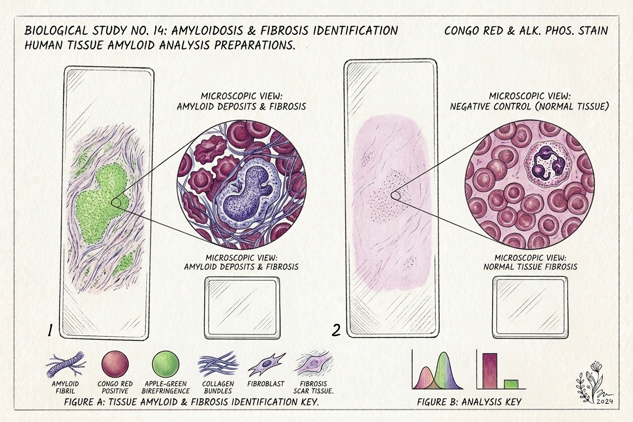
Congo red is one of the oldest amyloid stains and still the standard in pathology. When ordered amyloid fibrils bind the dye and are viewed between crossed polarizers, they show the well-known apple-green birefringence. Pathologists use it on biopsy tissue when they suspect systemic amyloidosis.
It has well-documented pitfalls. A 2019 historical review in Bioscience Reports concluded that Congo red staining alone isn't enough to confirm that aggregates are amyloid in the lab, or to diagnose amyloidosis in tissue. In clinical practice, a positive stain is followed by typing the protein, often with mass spectrometry, which we cover in types of amyloid tests.
Electron microscopy, X-ray diffraction and cryo-EM
These methods look at the structure directly, which makes them the most convincing, and the least practical for routine testing.
Electron microscopy shows fibrils as long, thin, typically unbranched threads. Scanning electron microscopy has also been used to look at the dense, matted fibrin in microclot research, as in the spike protein S1 and fibrin study from the Pretorius group.
X-ray fibre diffraction is how the cross-beta pattern was established. The 1997 study above remains a reference point.
Cryo-EM now resolves fibrils at near-atomic detail. A 2017 Nature study used it to solve the structure of tau filaments taken from an Alzheimer's brain. These methods need purified material, specialist instruments and expert analysis, so you won't find them on a lab requisition form.
What are seeding assays like RT-QuIC?
Some amyloids and prions can template, or "seed," their shape onto normal copies of the same protein. Seeding assays use that property to amplify tiny amounts. You mix the sample with normal protein, shake it in cycles and watch ThT fluorescence rise if seeds are present.
Real-time quaking-induced conversion (RT-QuIC) is the best-known example. In the 2011 Nature Medicine paper that introduced it, the assay detected 1 femtogram or more of abnormal prion protein in diluted brain tissue, and in a blinded set of 30 cerebrospinal fluid samples it reached greater than 80% sensitivity and 100% specificity for Creutzfeldt-Jakob disease. A 2021 Lancet Neurology review called RT-QuIC a major breakthrough for confident diagnosis of sporadic CJD before death, and updated diagnostic criteria include it. Related seeding assays are being studied for other neurodegenerative diseases.
Cross-seeding, where one protein's fibrils seed a different protein, is an active research question for spike protein. Our article on spike amyloid cross-seeding with Alzheimer's and prion proteins walks through those studies.
Antibodies for specific amyloid forms
Some antibodies recognize a shape rather than a sequence. A 2003 Science study found that soluble amyloid oligomers from different proteins share a common conformation that one antibody could recognize, distinct from mature fibrils, and that the antibody blocked their toxicity in cell experiments.
These tools matter because dyes like ThT and Congo red bind fibrils well but can miss smaller oligomers. Most conformation-specific antibodies are still research reagents. The clinical Alzheimer's blood tests are different: they measure amounts of particular peptides, like amyloid-beta 42/40 and p-tau217, not fibril structure.
Why researchers combine several methods
No single amyloid fibril test settles the question by itself.
Dyes are fast and cheap but can be fooled. Microscopy shows shape but samples only a small area. Structural methods are definitive but need purified material and specialist equipment. Seeding assays are extremely sensitive, which also makes them prone to contamination if handled carelessly.
So the usual pattern is to screen with a dye, confirm shape with microscopy, and, where it matters, pin down the structure with diffraction or cryo-EM. A finding that passes three different tests is far more convincing than one bright fluorescence reading. The spike protein studies below are a clear example of that layering.
How ThT is used to study spike protein fibrils
Spike protein amyloid research is a good example of stacking methods. In a 2022 Journal of the American Chemical Society study, Sofie Nyström and Per Hammarström screened the spike protein and found seven amyloid-prone sequences. Three 20-amino-acid peptides met three separate criteria: ThT kinetics typical of fibril growth, Congo red positivity and fibrillar shape under electron microscopy.
The full, folded spike protein didn't form fibrils on its own. It did form branching amyloid-like fibrils after being cut by neutrophil elastase, an enzyme released at inflamed sites. We explain that study step by step in how neutrophil elastase turns spike protein into amyloid.
A 2025 follow-up in Biochemistry tested seven spike amyloid fibrils with purified clotting proteins. Five didn't substantially interfere with clotting or clot breakdown. Fibrils from one fragment (Spike685) made fibrin clots markedly resistant to plasmin, while another (Spike601) bound up fibrinogen and slowed clot formation. These are test-tube results, and they don't show how often this happens in people.
How is ThT applied to blood?
Here's where amyloid fibril methods meet clinical questions. Pretorius, Kell and colleagues add ThT to platelet-poor plasma and image it with a fluorescence microscope. In their 2022 Biochemical Journal review, they describe fibrin amyloid microclots as fairly easy to measure this way.
The same group later acknowledged that manual microscopy lacked objectivity and throughput, and in a 2023 Heliyon paper it showed that imaging flow cytometry could count and size ThT-positive microclots more objectively. Neither approach has an agreed clinical cutoff yet. Our pillar on the amyloid fibrin test covers what that means for patients, including how these results compare with D-dimer, and our fibrin amyloid microclots research summary covers the wider evidence.
Clinical next step
Curious what Thioflavin T shows in your blood?
Our at-home amyloid fibrin test applies ThT fluorescence microscopy to a dried blood sample and returns a shareable video report.
Start screeningWhere our kit fits
Our at-home amyloid test applies one method from this list, Thioflavin T fluorescence microscopy, to a finger-prick dried blood sample. The sample goes to our partner laboratory in Mexico City, where it's stained with ThT and recorded as 6K 12-bit RAW video microscopy at 20× magnification under UV illumination. The footage is reviewed with AI-assisted fibril detection plus trained human oversight, and you get a Videographic Microscopy Report in a secure portal, usually within 1 day of the lab receiving the sample (5–7 business days from kit delivery including shipping). It costs $160 USD (3,200 MXN).
Precision matters here. ThT microscopy shows beta-sheet amyloid structure in the sample; it doesn't identify the protein, and it doesn't measure spike protein. The kit isn't a diagnostic test or a medical device, isn't an Alzheimer's test and isn't a test for systemic amyloidosis. The report is observational laboratory data for you to share with a qualified healthcare provider.
Which amyloid fibril test is right for your question?
Match the method to the question:
- "Do I have systemic amyloidosis?" That's a tissue biopsy with Congo red and protein typing, ordered by a specialist.
- "Do I have Alzheimer's pathology?" That's plasma p-tau217 and Aβ42/40 tests, amyloid PET or spinal fluid testing, not fibril stains.
- "Is there suspected prion disease?" That's RT-QuIC on spinal fluid, through a neurologist.
- "Are there fibrin amyloid microclots in my blood?" That's ThT microscopy or flow cytometry, a research-stage method.
Our guide to the types of amyloid tests lays these out in one table. If microclots are your main concern, continue with the amyloid fibrin test pillar and our explainer on amyloid microclots and Long COVID.
Frequently asked questions
What is the gold standard test for amyloid fibrils?
It depends on the setting. In a lab, structure-level methods such as X-ray fibre diffraction and cryo-electron microscopy give the most definitive picture of the cross-beta fold. In hospital pathology, Congo red staining of a biopsy, viewed under polarized light, is the standard first step for amyloidosis, followed by typing the protein. Thioflavin T is often called a gold standard for staining, but it works best alongside other checks.
Can Thioflavin T give a false positive?
Yes, it can. ThT fluoresces when it binds beta-sheet grooves, and other structures or compounds can sometimes increase or quench its signal. That's why careful studies don't rely on ThT alone. They confirm with Congo red, electron microscopy or structural methods, as the 2022 spike protein amyloid study did. In blood work, controls and consistent imaging settings help separate real signal from background.
Is there a blood test for amyloid fibrils?
Not in the sense of one validated clinical test for all fibrils. Alzheimer's blood tests measure soluble amyloid-beta and p-tau levels, not fibrils. Systemic amyloidosis is confirmed in tissue, not blood. Research groups use Thioflavin T microscopy and flow cytometry to look for fibrin amyloid microclots in plasma, and some labs offer that commercially as observational data, but it isn't a standardized diagnostic test.
What's the difference between an amyloid fibril and an amyloid oligomer?
Fibrils are long, ordered, insoluble structures that dyes like ThT and Congo red bind well. Oligomers are smaller, soluble clusters that form along the way and may be more toxic in some diseases. A 2003 Science study found that soluble oligomers from different proteins share a common structure that a specific antibody could recognize, distinct from fibrils. Fibril stains often miss oligomers.
Do spike protein amyloid fibrils show up on a fibrin microclot test?
A ThT-based microclot test sees beta-sheet amyloid structure, not which protein made it. Lab studies show spike protein fragments can form ThT-positive fibrils and that some can make fibrin clots resist breakdown. A blood smear image can't tell spike fibrils apart from fibrin amyloid, and it doesn't measure spike protein. That needs a spike-specific assay.
Sources
- Molecular mechanism of Thioflavin-T binding to amyloid fibrils. Biochimica et Biophysica Acta, 2010
- Congo Red and amyloids: history and relationship. Bioscience Reports, 2019
- Common core structure of amyloid fibrils by synchrotron X-ray diffraction. Journal of Molecular Biology, 1997
- Cryo-EM structures of tau filaments from Alzheimer's disease. Nature, 2017
- Ultrasensitive human prion detection in cerebrospinal fluid by real-time quaking-induced conversion. Nature Medicine, 2011
- Biomarkers and diagnostic guidelines for sporadic Creutzfeldt-Jakob disease. The Lancet Neurology, 2021
- Common structure of soluble amyloid oligomers implies common mechanism of pathogenesis. Science, 2003
- Amyloidogenesis of SARS-CoV-2 Spike Protein. Journal of the American Chemical Society, 2022
- SARS-CoV-2 Spike Protein Amyloid Fibrils Impair Fibrin Formation and Fibrinolysis. Biochemistry, 2025
- A central role for amyloid fibrin microclots in long COVID/PASC: origins and therapeutic implications. Biochemical Journal, 2022
- Accelerating discovery: A novel flow cytometric method for detecting fibrin(ogen) amyloid microclots using long COVID as a model. Heliyon, 2023
At-Home Amyloid Test
Curious what Thioflavin T shows in your blood?
Our at-home amyloid fibrin test applies ThT fluorescence microscopy to a dried blood sample and returns a shareable video report.
Next steps
- Amyloid Fibrin Test: How Fibrin Amyloid Microclots Are DetectedWhat an amyloid fibrin test is, how Thioflavin T microscopy spots fibrin amyloid microclots, how it differs from D-dimer, and what it can't tell you.
- Types of Amyloid Tests: Blood Tests, PET, CSF, Biopsy and MicroclotsCompare the types of amyloid tests: Alzheimer's blood tests, amyloid PET, spinal fluid, amyloidosis biopsy and fibrin amyloid microclot microscopy.
- Spike Protein Amyloid Fibrils: How Spike685 Makes Clots Resist LysisHow spike protein amyloid fibrils change fibrin clots: what the 2025 Linköping study found for Spike601 and Spike685, and what it cannot yet tell us.

Tim Ottowitz
Founder & Research Lead
Tim Ottowitz founded Amyloid Test and writes its Science Hub, turning fibrin amyloid and spike protein research into plain-language guides that stay close to the published evidence.
Keep reading
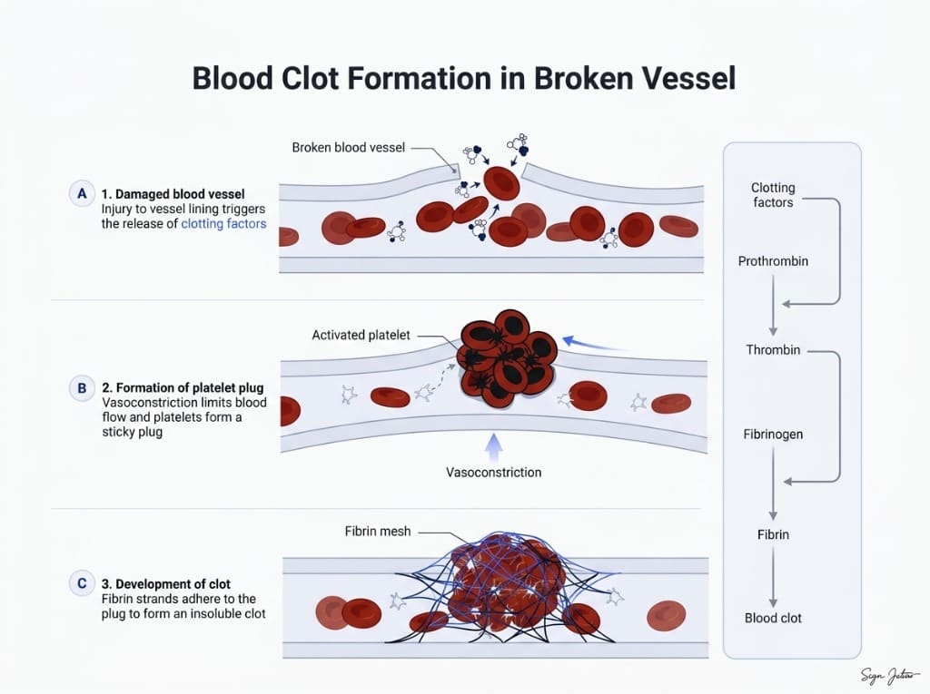
D-Dimer vs Microclot Test: Why a Normal D-Dimer Can Miss Microclots
D-dimer vs microclot test compared: what each measures, why D-dimer can read normal in Long COVID, and how Thioflavin T microscopy finds fibrin amyloid.
Neutrophil Elastase and Spike Protein: How Amyloid Forms
Neutrophil elastase cuts SARS-CoV-2 spike into amyloid-forming fragments. A plain-English walkthrough of the 2022 JACS study and what it does not show.
Spike Protein Antibody vs Antigen Testing: What Each One Measures
Spike antibody tests measure immune memory, not spike protein. What antigen testing adds, why detox protocols stall, and what a serious panel includes.