Amyloid Fibrin Test: How Fibrin Amyloid Microclots Are Detected
Short answer
An amyloid fibrin test looks for fibrin amyloid microclots: small clots of misfolded fibrin that resist the body's normal clot-dissolving system. A blood sample is stained with Thioflavin T, a dye that fluoresces when it binds amyloid, and examined under a fluorescence microscope. It's a research-stage method, not a standardized diagnostic test, and it measures something different from D-dimer or routine clotting panels.
Key takeaways
- 1An amyloid fibrin test uses Thioflavin T fluorescence microscopy to look for fibrinolysis-resistant fibrin amyloid microclots in blood.
- 2The method comes from research by Resia Pretorius, Douglas Kell and colleagues, who reported these microclots in Long COVID plasma and in other inflammatory conditions.
- 3D-dimer measures fragments released when clots break down, so it answers a different question than a microclot test.
- 4There's no agreed clinical cutoff, and independent researchers have challenged the microclot hypothesis, so results are observational, not diagnostic.
- 5Any result is best reviewed with a clinician alongside your symptoms and standard tests.
If your routine bloodwork keeps coming back normal while you still feel unwell after COVID-19 or vaccination, you've probably run into the term "microclots." This guide is for you, and for the clinicians you'll talk to. By the end you'll know what an amyloid fibrin test looks for, how it works, how it differs from D-dimer, and where its limits are.
What is an amyloid fibrin test?
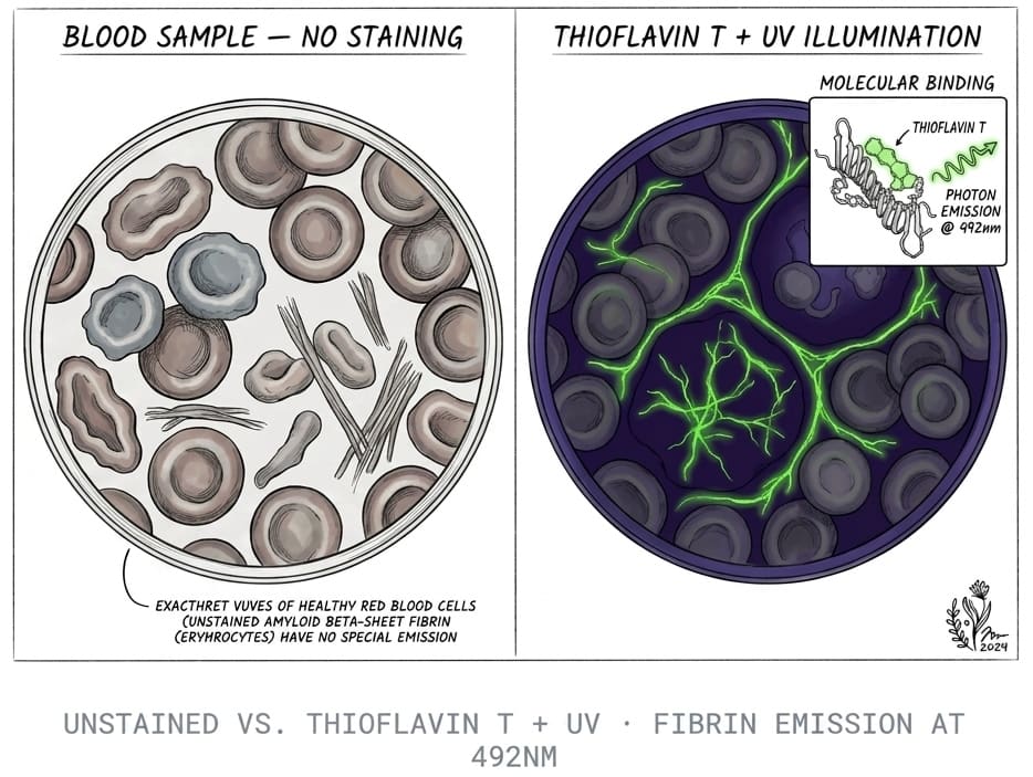
An amyloid fibrin test checks a blood sample for fibrin amyloid microclots, also called fibrinaloid microclots. These are small clumps of fibrin, the protein that forms the mesh of a normal blood clot, that have folded into an amyloid-like structure. The test stains the sample with Thioflavin T (ThT), a dye that lights up when it binds amyloid, and looks at it under a fluorescence microscope.
The idea comes from a research program led by Resia Pretorius at Stellenbosch University and Douglas Kell at the University of Liverpool. In a 2022 review in the Biochemical Journal, they described how fibrinogen can clot into an anomalous amyloid form that's relatively resistant to breakdown, and said these microclots are fairly easy to see in platelet-poor plasma with ThT and a simple fluorescence microscope. Our longer walkthrough of that body of work is in amyloid microclots research, and what microclots are is the plain-language starting point.
It's worth being clear on scope from the start. This isn't a test for Alzheimer's disease or for systemic amyloidosis, both of which use different methods. If you're trying to work out which test matches which question, our guide to the types of amyloid tests compares them side by side.
What are fibrin amyloid microclots?
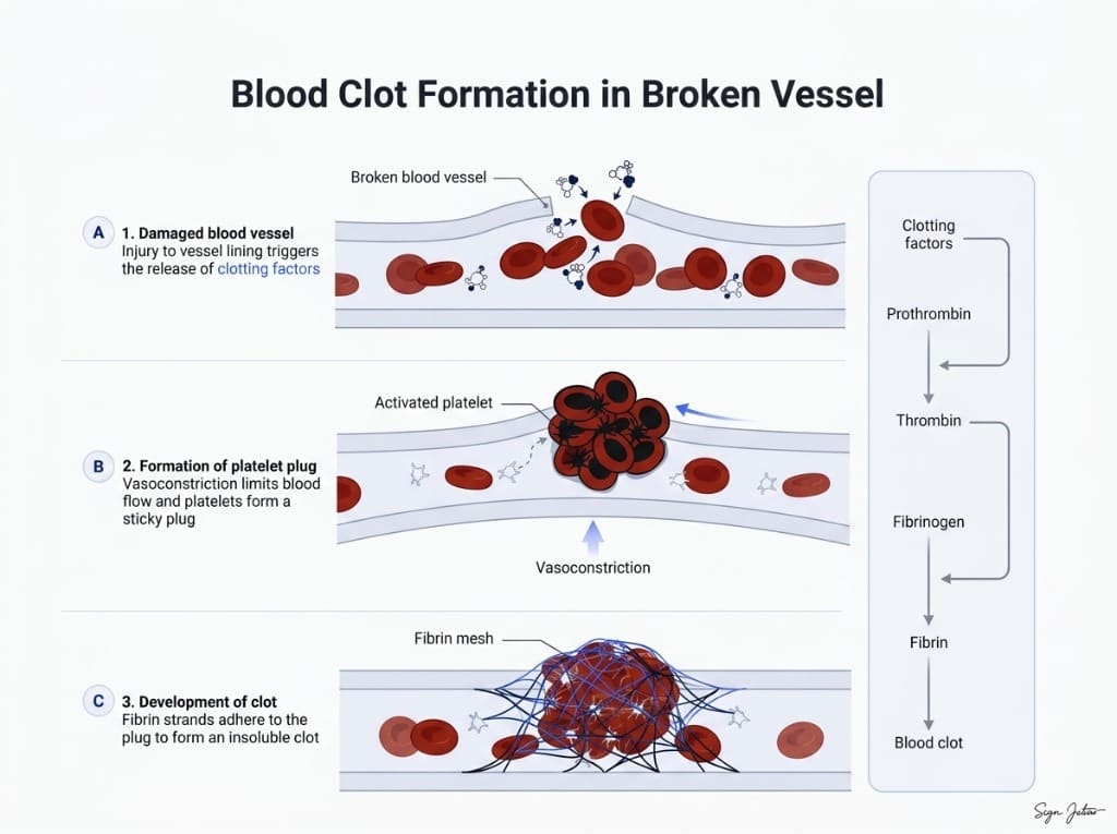
In a normal clot, the enzyme thrombin turns soluble fibrinogen into fibrin strands that knit together. Once the injury heals, the clot-dissolving system (fibrinolysis, driven mainly by the enzyme plasmin) breaks the mesh down again. That cycle happens constantly and is healthy.
Fibrin amyloid microclots behave differently. The Pretorius and Kell group reports that, under certain inflammatory triggers, fibrin forms denser, beta-sheet-rich deposits that resist plasmin. In their 2021 Cardiovascular Diabetology study, plasma from people with acute COVID-19 and Long COVID contained large anomalous amyloid deposits that stayed resistant to digestion, compared with plasma from healthy controls and people with type 2 diabetes. Trapped inside were raised levels of α2-antiplasmin (a natural brake on clot breakdown), fibrinogen chains and serum amyloid A.
A follow-up proteomics study of 99 Long COVID patients and 29 controls found more molecules caught inside the microclots, including von Willebrand factor, platelet factor 4 and antibodies. For a plain-language overview of how this ties into symptoms, see amyloid microclots and Long COVID.
Why "amyloid"?
Amyloid isn't one protein. It's a shape: proteins stacked into long fibrils held together by beta-sheets. Many unrelated proteins can adopt it, from amyloid-beta in the brain to light chains in amyloidosis.
Calling these clots "fibrin amyloid" means fibrin has taken on that stacked, dye-binding structure. Our companion pillar on the amyloid fibril test covers how scientists confirm that structure in general.
How does an amyloid fibrin test work?
The research protocol is short on paper. Blood is spun down to platelet-poor plasma, ThT is added, and the sample is imaged with a fluorescence microscope. ThT on its own barely glows, but when it binds the beta-sheet grooves in amyloid it fluoresces brightly, so amyloid deposits stand out against a dark background.
What you look at afterward is the hard part. Researchers judge the number, size and spread of fluorescent deposits. The Pretorius group uses a published grading system for microclots and platelet pathology, and reported significant microclots and platelet changes in all 80 Long COVID samples it analyzed in that 2022 study.
Manual microscopy has known weak points, and the researchers themselves have said so. In a 2023 Heliyon paper, the same group wrote that its earlier microscopy approach lacked statistical strength, objectivity and fast throughput, and tested imaging flow cytometry to count and size microclots more objectively. They noted that differences between Long COVID and control samples showed up even with a 20× objective.
Microscopy vs flow cytometry
| Approach | What it measures | Strengths | Limits |
|---|---|---|---|
| ThT fluorescence microscopy | Fluorescent amyloid deposits seen in a plasma or blood smear | Simple equipment; you can see the shapes directly | Judgment-based grading; field selection matters; no clinical cutoff |
| Imaging flow cytometry with ThT | Count, size and brightness of particles flowing past a camera | More objective counting; analyzes the whole sample | Specialized instruments; still a research method |
| Proteomics of digested microclots | Which proteins are trapped inside | Explains what the clots contain | Expensive, slow, research labs only |
How is it different from D-dimer and standard clotting tests?
This is the question most people arrive with. MedlinePlus describes D-dimer as a protein fragment made when a blood clot dissolves. Doctors use it mainly to help check for deep vein thrombosis, pulmonary embolism and disseminated intravascular coagulation (DIC).
So D-dimer reports on clot breakdown. An amyloid fibrin test looks for clot material that the research describes as resisting breakdown. The Kruger proteomics paper makes the related point that standard soluble markers such as CRP may look normal, and that current biomarkers can't detect the molecules trapped inside microclots. That's a hypothesis about why routine labs can miss something, not proof that a normal D-dimer hides microclots.
| Test | What it measures | Main clinical use | Sample | Status |
|---|---|---|---|---|
| D-dimer | Fibrin fragments released as clots dissolve | Helping rule out DVT, PE; monitoring DIC | Venous blood | Standard, validated |
| PT/INR, aPTT | How fast plasma clots | Bleeding risk, warfarin and heparin monitoring | Venous blood | Standard, validated |
| Fibrinogen | Amount of clotting protein in plasma | Bleeding disorders, DIC, inflammation | Venous blood | Standard, validated |
| Amyloid fibrin test (ThT microscopy) | Fluorescent fibrin amyloid microclots | Research into Long COVID and inflammatory disease | Plasma or blood sample | Research-stage, no agreed cutoff |
Our detailed comparison, D-dimer vs microclot test, goes further into why the two can disagree.
Who is an amyloid fibrin test for?
Most people who look into it fall into a few groups:
- People with Long COVID whose fatigue, brain fog or breathlessness persists while standard tests look normal.
- People with persistent symptoms after vaccination. A small 2026 proteomics study by the Pretorius group compared microclot deposits from 14 people with post-vaccination/post-infection syndrome and 16 controls and found changes in coagulation and immune proteins that only partly overlapped with earlier Long COVID data. Our article on post-vaccination microclots and fibrinolysis covers the background.
- Researchers and clinicians tracking microclots in other inflammatory conditions, which the group has also studied.
It's probably not useful if you need to rule out an acute clot. Sudden leg swelling, chest pain or sudden breathlessness need emergency care and standard tests like D-dimer and imaging, not a microscopy kit.
What the results can and can't show
A microscopy report shows what was seen in the sample: whether ThT-positive deposits were present, roughly how many, how large, and how they were distributed. Video or image capture lets another person look at the same fields rather than relying on a single reader's summary.
What it can't show matters just as much:
- It can't tell you what caused the deposits. The research links them to infection, spike protein, inflammatory molecules and chronic disease, and a sample doesn't carry a label.
- It doesn't measure spike protein. That needs a spike-specific assay, which we discuss in spike protein persistence testing.
- It doesn't give a diagnosis or say whether symptoms come from microclots.
What does spike protein have to do with it?
Lab studies suggest a possible link. In a 2021 Bioscience Reports study, adding isolated spike protein S1 to healthy plasma changed the structure of fibrin(ogen) and made it more resistant to digestion. That was an in vitro experiment, not a measurement in patients. Our explainer on spike protein amyloid fibrils and fibrinolysis covers the newer lab work from Linköping on how spike fragments themselves can form amyloid.
Limitations you should know about
We'd rather you hear these from us than discover them later.
- It isn't standardized. There's no agreed reference range, no validated cutoff and no regulatory approval for microclot microscopy as a diagnostic test. The 2023 Heliyon paper opens by noting there are no established diagnostic tools to identify which Long COVID patients might benefit from specific treatments.
- The hypothesis is contested. In 2024, Beverley Hunt and colleagues published a critical appraisal of the microclot hypothesis. They concluded the inferences weren't based on evidence, that unlicensed antithrombotic use wasn't justified, and that apheresis shouldn't be offered outside well-designed trials. Kell, Khan and Pretorius published a reply defending the evidence. Both are worth reading.
- Most data come from one research network. Independent replication with blinded readers and larger control groups is still limited.
- Interests exist. The 2022 review discloses that Pretorius is a named inventor on a patent application covering fluorescence methods for microclot detection in Long COVID. That doesn't make the work wrong, but you should know it.
- Sample handling matters. Published protocols use platelet-poor plasma from a venous draw. Other formats, including dried finger-prick samples, are a different sample type, which is one more reason to read results as observational.
Clinical next step
Want to see what's in your own blood sample?
Our at-home amyloid fibrin test uses Thioflavin T fluorescence microscopy and delivers a Videographic Microscopy Report you can share with your doctor.
Start screeningHow can you get an amyloid fibrin test?
There are three routes, and they suit different people.
Through a clinic. Some Long COVID and integrative clinics offer microclot imaging as part of an assessment, usually alongside standard bloodwork. Our directory of microclot testing labs and treatment centers lists options by region.
Through a research study. University groups recruit participants from time to time. This usually means a venous blood draw and no individual clinical report.
With an at-home kit. Our at-home amyloid fibrin test is built for people who want data without a clinic visit. Here's exactly what it is:
- You collect a finger-prick dried blood sample at home and mail it to our partner laboratory in Mexico City.
- The lab stains it with Thioflavin T, the same staining principle used in the published Pretorius and Kell microclot research, and records 6K 12-bit RAW video microscopy at 20× magnification under UV illumination.
- Footage is reviewed with AI-assisted fibril detection plus trained human oversight.
- You get a Videographic Microscopy Report in a secure client portal and can share it with any healthcare provider.
- The report is ready within 1 day of the lab receiving your sample. From kit delivery to report, it typically takes 5–7 business days including shipping.
- The price is $160 USD (3,200 MXN), and it includes access to a directory of doctors experienced with amyloid-related care.
What it isn't: it's not a diagnostic test or a medical device. It provides laboratory observational data for informational and research purposes, and clinical interpretation belongs to a qualified healthcare provider. It doesn't measure spike protein, D-dimer, fibrinogen or any standard coagulation value. It isn't an Alzheimer's test and doesn't test for systemic amyloidosis.
What should you ask before ordering any microclot test?
Labs and clinics describe microclot testing in very different ways, so a few questions help you compare like with like:
- What sample do they use? Platelet-poor plasma from a venous draw, whole blood, or a dried finger-prick sample are different materials, and results aren't directly comparable across them.
- What stain and imaging method? Thioflavin T fluorescence microscopy and imaging flow cytometry are the methods in the published research. Ask which one, and at what magnification.
- Who reads the images, and how? Manual grading, software-assisted counting and human review each have trade-offs. Ask whether you get the images or video, or only a summary score.
- What does the report claim? Be wary of any service that calls a microclot result a diagnosis, ties it to a specific treatment, or quotes accuracy figures without a published study behind them.
- Can you share it? A report is only useful if your doctor can see it and understand what was done.
These questions apply to our own kit too. We've answered them for our kit in the section above, so you can hold us to the same standard.
Preparing for the test
Follow the collection instructions that come with whatever test you use, and write down the date, any recent illness, and your current medicines and supplements. Don't stop prescribed anticoagulants or antiplatelet drugs to "get a clearer result." That decision belongs to the clinician who prescribed them, and stopping can be dangerous.
What to do with a result
Bring it to a clinician, together with your symptom history and your standard labs. A result can support a conversation about next steps. It can't settle a diagnosis on its own, and nobody should start, stop or change anticoagulants based on a microscopy image alone.
If you're weighing approaches that are being studied, our review of nattokinase and lumbrokinase for microclots sets out what the lab and clinical evidence does and doesn't show. Our doctor directory can help you find someone familiar with this research. For the bigger day-to-day picture, the Long COVID survival guide covers pacing, tracking and red-flag symptoms.
Where to go next
If you want to understand the dye and the chemistry behind the method, read our pillar on the amyloid fibril test, which compares Thioflavin T, Congo red, electron microscopy and seeding assays. If you're still sorting out which amyloid test matches your question, start with types of amyloid tests.
Frequently asked questions
Is an amyloid fibrin test the same as an Alzheimer's amyloid test?
No. Alzheimer's amyloid tests look for amyloid-beta plaques in the brain, using plasma Aβ42/40 and p-tau217 blood tests, amyloid PET scans or spinal fluid. An amyloid fibrin test looks at fibrin, the clotting protein, in a blood sample and checks whether it has formed amyloid-like microclots. The two share the word amyloid but measure unrelated things and answer different medical questions.
Can a normal D-dimer rule out microclots?
Not by itself. D-dimer is a protein fragment released when a clot is broken down, and it's mainly used to help rule out deep vein thrombosis, pulmonary embolism and DIC. Fibrin amyloid microclots are described in the research as resistant to breakdown, so a normal D-dimer doesn't tell you whether they're present. It also doesn't prove they are, which is why both results need clinical context.
Do I need to stop blood thinners or supplements before testing?
Don't stop or change any prescribed medication to prepare for a test without talking to the doctor who prescribed it. Anticoagulants and antiplatelet drugs are prescribed for specific risks, and stopping them can be dangerous. If you're taking enzymes like nattokinase or a blood thinner, note it when you share your results so whoever reviews them can factor it in.
How accurate is an amyloid fibrin test?
There's no published accuracy figure for microclot microscopy against an agreed reference standard, because no reference standard exists yet. The research group behind the method has said its earlier microscopy lacked statistical strength and objectivity, and it has tested imaging flow cytometry to make counting more objective. Treat any result as observational data rather than a pass or fail diagnosis.
Who orders an amyloid fibrin test?
Most people who look into it have persistent symptoms after COVID-19 infection or vaccination, often with normal routine bloodwork. Some integrative and Long COVID clinics use microclot imaging in their workup, and some people order an at-home kit to bring data to their own doctor. It isn't part of standard guidelines, so many physicians won't have seen one before.
Can an amyloid fibrin test tell me which treatment to use?
No. No clinical guideline links a microclot result to a specific treatment, and controlled trials of anticoagulants, enzymes or apheresis for microclots are still limited. A result can be one piece of information in a conversation with a clinician, but treatment decisions should rest on your full medical picture, not on a single research-stage test.
Sources
- A central role for amyloid fibrin microclots in long COVID/PASC: origins and therapeutic implications. Biochemical Journal, 2022
- Persistent clotting protein pathology in Long COVID/Post-Acute Sequelae of COVID-19 (PASC) is accompanied by increased levels of antiplasmin. Cardiovascular Diabetology, 2021
- SARS-CoV-2 spike protein S1 induces fibrin(ogen) resistant to fibrinolysis: implications for microclot formation in COVID-19. Bioscience Reports, 2021
- Prevalence of symptoms, comorbidities, fibrin amyloid microclots and platelet pathology in individuals with Long COVID/PASC. Cardiovascular Diabetology, 2022
- Proteomics of fibrin amyloid microclots in long COVID/PASC shows many entrapped pro-inflammatory molecules that may also contribute to a failed fibrinolytic system. Cardiovascular Diabetology, 2022
- Accelerating discovery: A novel flow cytometric method for detecting fibrin(ogen) amyloid microclots using long COVID as a model. Heliyon, 2023
- Challenging the current hypothesis that thrombosis is responsible for the post-COVID-19 condition. Research and Practice in Thrombosis and Haemostasis, 2024
- Fibrinaloid microclots in long COVID: assessing the actual evidence properly. Research and Practice in Thrombosis and Haemostasis, 2024
- Proteomic signatures of COVID-19 Post-Vaccination/Post-Infection Syndrome (PV/PIS): insights into immune dysregulation and coagulopathy. Frontiers in Cellular and Infection Microbiology, 2026
- D-dimer Test. MedlinePlus, U.S. National Library of Medicine, 2024
At-Home Amyloid Test
Want to see what's in your own blood sample?
Our at-home amyloid fibrin test uses Thioflavin T fluorescence microscopy and delivers a Videographic Microscopy Report you can share with your doctor.
Next steps
- Amyloid Fibril Test: Methods Used to Detect Amyloid FibrilsHow an amyloid fibril test works: Thioflavin T, Congo red, electron microscopy, X-ray diffraction and seeding assays compared, with which are clinical.
- Types of Amyloid Tests: Blood Tests, PET, CSF, Biopsy and MicroclotsCompare the types of amyloid tests: Alzheimer's blood tests, amyloid PET, spinal fluid, amyloidosis biopsy and fibrin amyloid microclot microscopy.
- What Are Microclots? A Plain-Language Guide to Amyloid MicroclotsWhat microclots are, how fibrin folds into amyloid microclots that resist breakdown, the symptoms researchers link to them, and how they're tested.

Tim Ottowitz
Founder & Research Lead
Tim Ottowitz founded Amyloid Test and writes its Science Hub, turning fibrin amyloid and spike protein research into plain-language guides that stay close to the published evidence.
Keep reading
D-Dimer vs Microclot Test: Why a Normal D-Dimer Can Miss Microclots
D-dimer vs microclot test compared: what each measures, why D-dimer can read normal in Long COVID, and how Thioflavin T microscopy finds fibrin amyloid.
How to Test for Microclots: Methods, Options and What Results Mean
How to test for microclots: the methods researchers use, why D-dimer can't detect them, where to get a microclot test and how to read the result.
Amyloid Microclots: What the Research Shows So Far
What studies show about amyloid microclots in Long COVID: how fibrin forms them, why they resist breakdown, the key findings and the open questions.