Microclots Symptoms: Capillary Occlusion, Fatigue, and Dyspnea
English Narration
At-Home Blood Spot • 6K Video Microscopy • $160
Short answer
Microclot symptoms stem from microvascular hypoperfusion, where amyloid fibrin aggregates physically obstruct 5 to 10 micrometer capillary networks. When red blood cells cannot traverse blocked microvessels, tissues starve of oxygen despite normal pulse oximeter readings. Common symptoms include debilitating fatigue, unrefreshing sleep, post-exertional malaise, cognitive impairment, and air hunger that routine hospital laboratory tests consistently miss.
Key takeaways
- 1Microclot symptoms arise from microvascular hypoperfusion rather than systemic large-vessel blockages.
- 2Pulse oximeters often read 98% to 100% while peripheral tissues remain chronically hypoxic at the capillary level.
- 3Post-exertional malaise occurs when oxygen delivery fails to match cellular metabolic demand during physical or mental activity.
- 4Standard blood panels, D-dimer, and chest X-rays do not show microclots because amyloid cross-beta structures resist fibrinolysis.
- 5Fluorescence microscopy with Thioflavin T provides objective visual confirmation of anomalous fibrin aggregates.
One of the most frustrating experiences for patients with post-viral syndromes is having debilitating physical symptoms while standard lab work returns completely normal. Doctors run routine complete blood counts, cardiac enzymes, and metabolic panels, only to announce that everything appears fine.
This contradiction happens because conventional medical tests check for large-vessel clotting, overt organ failure, and gross cellular abnormalities. They were never designed to detect anomalous microvascular pathology.
When fibrinogen misfolds into amyloid sheets, it forms microclots measuring 5 to 50 micrometers. These microscopic deposits lodge in capillaries throughout the body, obstructing oxygen delivery and creating a distinct clinical picture known as microvascular hypoperfusion.
The Underlying Mechanism: Microvascular Hypoperfusion
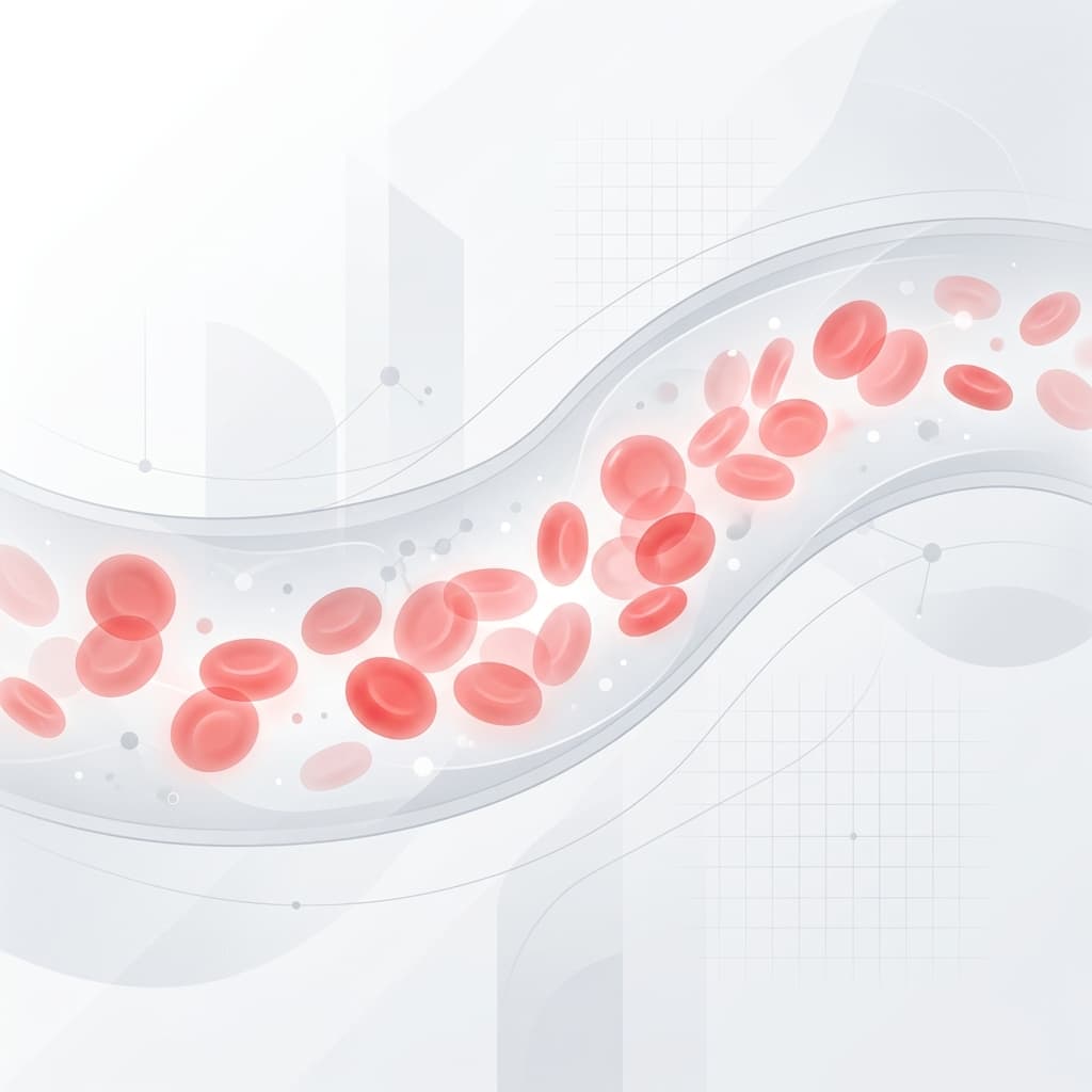
To understand microclots symptoms, you have to understand capillary diameter. Human capillaries are tiny: typically 5 to 8 micrometers across. A healthy red blood cell measures about 7.5 micrometers in diameter, meaning it must deform and squeeze single-file through capillary channels to deliver oxygen to adjacent cells.
When inflammatory stimuli or viral spike proteins trigger anomalous clotting, fibrinogen misfolds into amyloid aggregates. As explained in our scientific guide on what microclots are, these structures form rigid cross-beta sheets that resist natural enzymatic breakdown.
These insoluble aggregates circulate and physically wedge inside narrow capillary bifurcations. Red blood cells back up behind the obstruction. Surrounding tissues, whether in the brain, heart, lungs, or skeletal muscle, become starved of oxygen and nutrients.
The result is local tissue hypoxia and cellular starvation, occurring even when overall blood volume and central blood pressure remain completely normal.
Primary Microclots Symptoms Across Organ Systems
Because capillaries supply virtually every tissue in the human body, capillary occlusion produces multi-system symptoms that often appear unrelated on the surface.
1. Chronic Fatigue and Post-Exertional Malaise (PEM)
Profound, unrefreshing fatigue is the hallmark symptom of systemic microclotting. Unlike everyday tiredness that improves with a night of good sleep, microvascular fatigue stems from mitochondrial oxygen starvation.
When skeletal muscle fibers cannot obtain adequate oxygen through occluded capillary beds, cellular metabolism shifts from aerobic respiration to inefficient anaerobic glycolysis. Lactic acid accumulates rapidly during minimal exertion.
Patients frequently experience severe post-exertional malaise (PEM), where modest physical or mental activities trigger a delayed symptom crash lasting days or weeks.
2. Cognitive Dysfunction and "Brain Fog"
The human brain consumes approximately 20% of the body's resting oxygen supply despite representing only 2% of total body mass. It contains hundreds of miles of fragile microvessels.
When amyloid microclots lodge in cerebral microvessels, local perfusion drops. Patients experience debilitating neurocognitive symptoms:
- Severe memory lapses and word-finding difficulties
- Inability to focus or sustain attention on basic tasks
- Spatial disorientation and mental processing delays
- Sensation of mental heaviness or internal head pressure
3. Dyspnea and "Air Hunger" with Normal Oxygen Saturation
Shortness of breath is common among individuals with persistent microclots. Patients often describe an agonizing sensation of "air hunger", feeling as though they cannot take a satisfying breath even when resting.
When evaluated at an urgent care or emergency clinic, their pulse oximeter consistently reads 98% to 100%, and chest X-rays appear unremarkable. This paradox confuses clinicians unfamiliar with microvascular pathology.
In the alveoli of the lungs, gas exchange occurs across a delicate capillary web. Microclots obstruct pulmonary capillaries, creating a ventilation-perfusion mismatch. Oxygen enters the lung sacs normally, but red blood cells cannot transit the blocked capillaries to pick it up in sufficient quantities, triggering dyspnea receptors.
4. Muscle Weakness and Exercise Intolerance
Skeletal muscles require a massive surge in capillary blood flow during movement. When microclots block capillary recruitment, muscles rapidly run out of fuel. Patients report heavy, leaden limbs, muscle twitching (fasciculations), and burning pain during ordinary activities like walking up a flight of stairs or carrying groceries.
5. Peripheral and Autonomic Symptoms
When terminal capillaries in the skin and extremities fail to perfuse, patients frequently report:
- Cold hands and feet, often with purple or mottled skin (livedo reticularis)
- Burning nerve pain and paresthesias in fingers and toes
- Orthostatic tachycardia and dizziness upon standing due to autonomic capillary pooling
Why Standard Blood Tests Miss Microclots
Patients frequently ask why routine hospital blood work does not alert their doctor to these clotting issues. The explanation lies in the distinction between soluble clotting markers and insoluble amyloid structures.
Standard coagulopathy panels measure the time it takes for liquid plasma to clot (PT/INR and PTT). As detailed in our breakdown of D-dimer vs microclot testing, standard D-dimer tests measure soluble crosslinked fragments generated when the enzyme plasmin successfully dissolves a fibrin clot.
Amyloid fibrin microclots carry a dense, hydrophobic cross-beta steric zipper structure. This structure physically blocks plasmin from binding, leaving the clots largely indissoluble.
Because plasmin cannot cleave these amyloid structures, D-dimer levels remain completely normal. A standard complete blood count measures suspended cellular counts, not optical morphology. To evaluate microvascular issues, specialized Long COVID blood testing protocols are necessary.
How Microclots Are Objectively Evaluated
Because routine biochemical assays cannot see microclots, researchers developed direct optical visualization techniques using fluorescence microscopy.
Thioflavin T (ThT) Fluorescence Staining
Thioflavin T is an amyloid-specific fluorophore. In liquid solution, ThT rotates freely and emits negligible light. When it binds into the narrow channels between beta-sheets in amyloid fibrils, its molecular rotation becomes immobilized, causing a dramatic 1,000-fold increase in fluorescent emission.
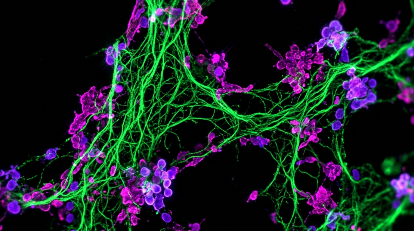
When human blood or plasma is stained with ThT and exposed to ultraviolet light, amyloid microclots glow with brilliant fluorescence under the microscope. Our guide on the amyloid fibrin test covers the complete laboratory methodology.
Visual Microscopic Staging
Experienced laboratories grade microclot presence using a four-stage morphological scale:
- Stage 1: Very few, tiny fluorescent deposits (<5 micrometers). Normal healthy baseline.
- Stage 2: Scattered microclots measuring 5 to 20 micrometers.
- Stage 3: Frequent, dense aggregates measuring 20 to 50 micrometers that obstruct fields of view.
- Stage 4: Massive, sheet-like amyloid mats exceeding 50 micrometers with extensive platelet trapping.
Next Steps for Patients Experiencing Symptoms
If you suspect your chronic symptoms are driven by persistent microclots, consider taking the following structured steps:
- Rule Out Acute Emergencies First: Always have a licensed medical provider rule out acute cardiovascular issues, pulmonary embolisms, structural lung diseases, and major metabolic disorders.
- Track Your Symptom Patterns: Note the specific triggers that cause post-exertional crashes or cognitive flares. Documenting these patterns helps distinguish microvascular hypoperfusion from other conditions.
- Seek Objective Microscopic Evidence: Rather than guessing whether your symptoms are caused by microvascular clotting, an at-home blood test provides high-definition 6K video fluorescence microscopy of your sample for $160 USD.
- Partner with a Knowledgeable Clinician: Share your visual microscopy report and peer-reviewed microclot literature with an open-minded healthcare practitioner to discuss supportive options.
Frequently asked questions
Why do pulse oximeters show normal oxygen when I feel short of breath?
A finger pulse oximeter measures arterial hemoglobin saturation in larger digital arteries before blood reaches terminal capillaries. When microclots clog small capillary beds in the lungs and skeletal muscle, red blood cells cannot exchange gas efficiently. You experience severe tissue hypoxia and air hunger even though the blood leaving your heart is 99% saturated.
What are the most common microclots symptoms reported by patients?
The most frequent symptoms include severe chronic fatigue, post-exertional malaise, cognitive dysfunction or brain fog, shortness of breath on mild exertion, muscle aches, temperature dysregulation, and cold or discolored extremities.
Can routine hospital lab work show microclots in blood?
No. Routine hospital lab tests like complete blood counts, metabolic panels, and D-dimer tests measure liquid blood dynamics and soluble protein breakdown fragments. Fibrin amyloid microclots are insoluble, cross-beta sheet structures that evade enzymatic lysis and require specialized fluorescence microscopy to visualize.
How does microclot testing help if my standard medical tests are all normal?
Normal lab panels often leave patients without clinical validation or clear direction. Visualizing Thioflavin T-positive amyloid microclots provides objective microscopic evidence of ongoing microvascular pathology, helping you and your physician explore targeted supportive protocols.
Sources
- A central role for amyloid fibrin microclots in long COVID/PASC: origins and therapeutic implications. Biochemical Journal, 2022
- Persistent clotting protein pathology in Long COVID/Post-Acute Sequelae of COVID-19 (PASC) is accompanied by increased levels of antiplasmin. Cardiovascular Diabetology, 2021
- Cardiovascular and haematological pathology in long COVID. Nature Reviews Cardiology, 2024
- Fibrinaloid microclots and systemic inflammation in post-acute sequelae of COVID-19. eBioMedicine, 2022
At-Home Amyloid Test
Are microclots driving your unexplained symptoms?
Order an at-home blood test kit for $160 USD. Receive 6K RAW video fluorescence microscopy and an objective laboratory report.
Next steps
- What Are Microclots? A Plain-Language Guide to Amyloid MicroclotsWhat microclots are, how fibrin folds into amyloid microclots that resist breakdown, the symptoms researchers link to them, and how they're tested.
- How to Test for Microclots: Methods, Options and What Results MeanHow to test for microclots: the methods researchers use, why D-dimer can't detect them, where to get a microclot test and how to read the result.
- Amyloid Fibrin Test: How Fibrin Amyloid Microclots Are DetectedWhat an amyloid fibrin test is, how Thioflavin T microscopy spots fibrin amyloid microclots, how it differs from D-dimer, and what it can't tell you.

Tim Ottowitz
Founder & Research Lead
Tim Ottowitz founded Amyloid Test and writes its Science Hub, turning fibrin amyloid and spike protein research into plain-language guides that stay close to the published evidence.
Keep reading
Amyloid Microclots: What the Research Shows So Far
What studies show about amyloid microclots in Long COVID: how fibrin forms them, why they resist breakdown, the key findings and the open questions.
Best at Home Microclot Test Kits in 2026: An Evidence-Based Guide
Compare at-home microclot test kits and clinical testing options. Learn how ThT staining, dried-blood stability, and turnaround times compare.
Anomalous Amyloid Microclots: Phenotypes and Phosphatidylserine
Explore the biophysics of anomalous amyloid microclots. Discover the 4 structural phenotypes, phosphatidylserine membrane flipping, and ThT microscopy.