Amyloid Microclots Blood Test: Methods, Costs, and Home Kits
English Narration
At-Home Blood Spot • 6K Video Microscopy • $160
Short answer
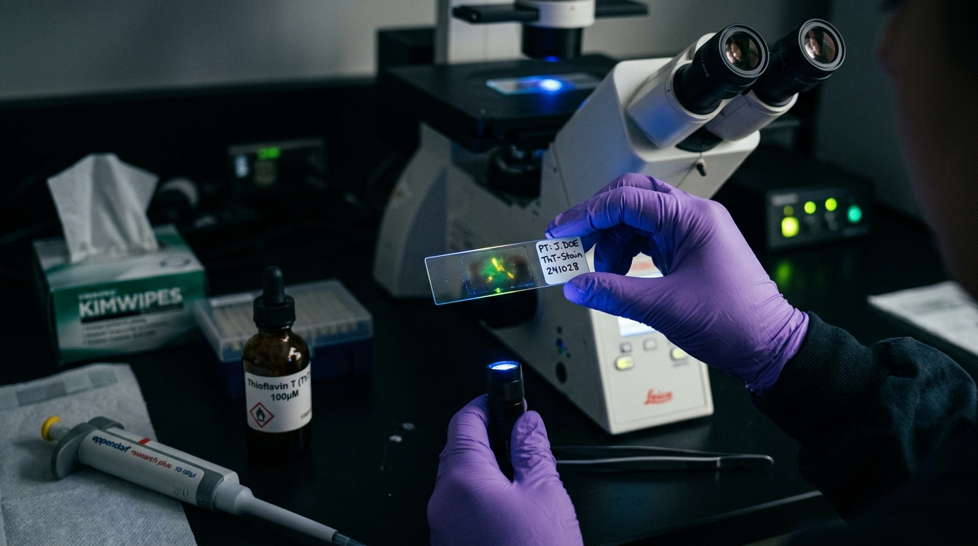
An amyloid microclots blood test uses Thioflavin T (ThT) fluorescent staining to detect anomalous cross-beta sheet fibrin aggregates under ultraviolet microscopy. Standard coagulation panels miss these structures because they resist enzymatic breakdown. While hospital testing remains largely confined to clinical trials, at-home finger-prick kits now provide accessible 6K video microscopy verification for $160 USD.
Key takeaways
- 1An amyloid microclots blood test visualizes physical protein folding rather than measuring liquid coagulation times.
- 2Thioflavin T dye binds specifically to cross-beta sheet spines in anomalous fibrin, causing them to fluoresce under UV light.
- 3Standard tests like D-dimer, PT/INR, and fibrinogen cannot detect amyloid microclots because cross-beta sheets resist enzymatic digestion.
- 4Clinical hospital access is severely constrained by a lack of FDA billing codes and automated diagnostic equipment.
- 5At-home dried blood collection provides an accessible testing option for $160 USD, delivering 6K RAW video microscopy.
Patients investigating chronic fatigue, brain fog, and vascular symptoms after viral infection frequently search for an amyloid microclots blood test. Yet when they ask their local healthcare provider or hospital laboratory, they are often met with blank stares.
This gap exists because microclot testing belongs to the frontier of translational hematology. While academic researchers have published dozens of papers documenting these structures, standard healthcare systems have not yet operationalized the test into routine clinical menus.
This comprehensive guide explains how an amyloid microclots blood test works, why standard hospital tests cannot replicate it, what laboratory options exist, and how at-home collection kits provide an accessible alternative.
The Chemistry Behind the Test: Thioflavin T Staining
To understand an amyloid microclots blood test, one must examine how the laboratory identifies anomalous clot material.
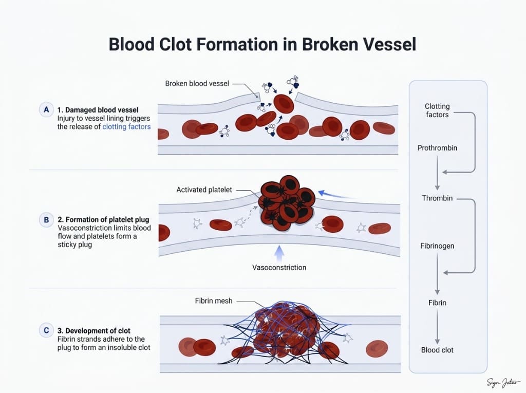
Under normal physiological conditions, thrombin cleaves soluble fibrinogen into fibrin monomers. These monomers spontaneously polymerize into a flexible, porous meshwork that seals vascular injuries. Once healing is underway, the enzyme plasmin degrades this fibrin mesh cleanly.
In inflammatory states and post-viral syndromes, fibrinogen undergoes an anomalous conformational change. As detailed in our comprehensive guide on the amyloid fibrin test, the protein misfolds into rigid cross-beta sheets known as amyloid fibrils.
Because these misfolded sheets carry tight, interlocking steric zippers, conventional optical microscopy cannot clearly differentiate them from regular cellular debris. Laboratories solve this problem using Thioflavin T (ThT).
Thioflavin T is a benzothiazole fluorophore dye. In aqueous solution, the dye molecules rotate freely, dissipating absorbed light as harmless vibrational energy.
When ThT molecules encounter cross-beta amyloid channels, they slide into the narrow grooves between stacked beta-sheets. The surrounding protein structure immobilizes the dye molecule.
When the microscopist illuminates the slide with ultraviolet excitation light (around 485 nm wavelength), the immobilized ThT molecules fluoresce brilliantly in the green-yellow spectrum (emission at 535 nm). The background remains completely dark, allowing the technician to visualize and quantify microscopic amyloid deposits with extreme optical contrast.
Why Routine Hospital Blood Panels Miss Microclots
Patients frequently ask: "If I have microclots in my blood, why did my routine hospital bloodwork come back clean?"
Conventional clinical hematology relies on automated biochemical analyzers designed to detect macrovascular thromboses or bleeding disorders:
- Complete Blood Count (CBC): Counts suspended cellular populations (red blood cells, white blood cells, platelets). It cannot assess whether platelets are entrapped in microscopic amyloid aggregates.
- Prothrombin Time (PT/INR) and PTT: Measures the speed of liquid clot formation in a test tube. It does not examine the physical structure or degradation resistance of the clot.
- D-Dimer Assay: As explained in our comparison of D-dimer vs microclot testing, D-dimer only measures fragments released when plasmin successfully dissolves a fibrin clot. Because amyloid cross-beta sheets physically resist plasmin binding, microclots persist without elevating D-dimer.
To learn more about the broader diagnostic landscape, read our overview of the types of amyloid tests.
Current Testing Routes: University Labs vs At-Home Kits
Because Quest Diagnostics and Labcorp do not offer microclot testing on commercial requisition sheets, patients have two primary avenues to obtain testing.
1. Academic Research Trials
The earliest testing was performed exclusively in academic research environments, notably Professor Resia Pretorius's laboratory at Stellenbosch University and affiliated research programs in Europe.
- Pros: Highest level of academic rigor and dual-staining protocols (e.g. CD62P platelet markers combined with ThT).
- Cons: Testing is tied strictly to active research study enrollment. Individual patient reports are rarely returned with actionable clinical turnaround.
2. Specialized In-Person Integrative Clinics
A limited number of private specialty practices offer microclot imaging as part of complex clinical workups.
- Pros: Includes direct clinical consultation and physical examination.
- Cons: Expensive ($800 to $2,500 total out-of-pocket), requires extensive travel for patients outside regional hubs, and involves multi-month waitlists.
3. At-Home Dried Blood Sample Kits
To eliminate the logistical and financial bottlenecks of clinical travel, at-home collection kits were developed.
Our at-home amyloid test costs $160 USD (3,200 MXN) all-inclusive. You collect a capillary blood spot via a single-use sterile lancet, allow the slide to air-dry, and return it in a prepaid mailer to our partner laboratory in Mexico City.
Dried blood collection solves a fundamental transport challenge. Liquid blood in vacuum tubes begins degrading within hours unless subjected to rapid centrifugation and frozen shipping on dry ice.
Because cross-beta amyloid fibrils possess remarkable thermodynamic and structural stability, dried blood spot samples preserve fibril morphology during postal transit without cold-chain requirements.
Upon receipt, the laboratory prepares the specimen with ThT staining and captures 6K 12-bit RAW video fluorescence microscopy at 20x optical magnification. The footage is reviewed using computer-assisted detection alongside trained human oversight, and the complete video microscopy report is delivered to your secure portal.
What Your Test Report Delivers
An objective amyloid microclots blood test report provides visual documentation rather than a generic pass/fail stamp:
- High-Definition Video Recordings: 6K RAW video sweeps of multiple high-power microscopic fields under ultraviolet excitation.
- Morphological Staging: Objective classification based on aggregate size (Stage 1 minimal under 5 microns up to Stage 4 confluent sheets exceeding 50 microns).
- Platelet Activation Observations: Visual assessment of platelet clumping and cellular debris entrapment within the fibrin network.
- Physician-Ready Documentation: Formatted files that you can download and share directly with your clinical care team.
Medical Status and Proper Context
It is essential to understand the clinical scope of microclot testing:
- Observational Research Assay: Microclot testing is an investigational laboratory assay, not an FDA-cleared diagnostic device.
- Not a Disease Diagnosis: The test demonstrates whether anomalous amyloid fibrin aggregates are present. It does not diagnose Long COVID, ME/CFS, or any specific illness.
- No Circulating Spike Protein Measurement: The test evaluates downstream fibrin morphology; it does not measure active circulating spike protein levels.
- Physician Partnership: Your report is designed to facilitate informed discussions with your medical provider, who can place the findings in the context of your complete medical history.
Frequently asked questions
Can I get an amyloid microclots blood test at Quest Diagnostics or Labcorp?
No. Major commercial diagnostic chains like Quest and Labcorp only run automated high-throughput biochemical analyzers. Because microclot analysis requires specialized fluorescence microscopy, manual staining with Thioflavin T, and optical grading by a trained microscopist, it is not available on standard commercial test menus.
How does this test differ from routine clotting tests?
Routine clotting tests measure how quickly liquid plasma clots or breaks down soluble fibrin fragments. An amyloid microclots blood test uses fluorescent dye to visually inspect blood for insoluble, misfolded amyloid fibrils that resist breakdown.
Is the test covered by private medical insurance?
No. Microclot testing is currently an investigational research assay without established American Medical Association CPT billing codes. As a result, commercial insurers, Medicare, and Medicaid do not cover the cost.
What is the turnaround time for at-home microclot testing?
After collecting a capillary finger-prick sample and mailing it to the laboratory, imaging is completed within 24 to 48 hours of specimen arrival. Total turnaround time, including international mail transit, typically ranges between 2 and 3 weeks.
Sources
- SARS-CoV-2 spike protein S1 induces fibrin(ogen) resistant to fibrinolysis. Biochemical Journal, 2021
- A central role for amyloid fibrin microclots in long COVID/PASC: origins and therapeutic implications. Biochemical Journal, 2022
- Persistent clotting protein pathology in Long COVID/Post-Acute Sequelae of COVID-19 (PASC) is accompanied by increased levels of antiplasmin. Cardiovascular Diabetology, 2021
At-Home Amyloid Test
Check your blood for persistent amyloid microclots
Order an at-home blood test kit for $160 USD. Receive 6K RAW video fluorescence microscopy and an objective laboratory report.
Next steps
- Amyloid Fibrin Test: How Fibrin Amyloid Microclots Are DetectedWhat an amyloid fibrin test is, how Thioflavin T microscopy spots fibrin amyloid microclots, how it differs from D-dimer, and what it can't tell you.
- Amyloid Fibril Test: Methods Used to Detect Amyloid FibrilsHow an amyloid fibril test works: Thioflavin T, Congo red, electron microscopy, X-ray diffraction and seeding assays compared, with which are clinical.
- Types of Amyloid Tests: Blood Tests, PET, CSF, Biopsy and MicroclotsCompare the types of amyloid tests: Alzheimer's blood tests, amyloid PET, spinal fluid, amyloidosis biopsy and fibrin amyloid microclot microscopy.

Tim Ottowitz
Founder & Research Lead
Tim Ottowitz founded Amyloid Test and writes its Science Hub, turning fibrin amyloid and spike protein research into plain-language guides that stay close to the published evidence.
Keep reading
At-Home Microclot Test vs Clinic: Cost, Labs, and Accuracy
Compare at-home microclot tests with in-person clinic testing. Examine cost, wait times, sample stability, and 6K ThT fluorescence microscopy methods.
D-Dimer vs Microclot Test: Why a Normal D-Dimer Can Miss Microclots
D-dimer vs microclot test compared: what each measures, why D-dimer can read normal in Long COVID, and how Thioflavin T microscopy finds fibrin amyloid.
Flow Clotometry vs Microscopy: How Labs Actually Count Microclots
Compare flow clotometry with 6K video fluorescence microscopy for microclots. Learn how sample prep, hydrodynamic shear, and optics affect test accuracy.