Spike Protein Amyloid Fibrils: How Spike685 Makes Clots Resist Lysis
Short answer
Spike protein amyloid fibrils are stable, stacked protein strands that short pieces of the spike protein, especially Spike685, form in lab experiments. In a 2025 test-tube study, clots containing Spike685 fibrils were dense and resisted plasmin, the clot-dissolving enzyme. No patients were studied, so this is a lab result, not proof of what happens in people.
English Narration
At-Home Blood Spot • 6K Video Microscopy • $160
Key takeaways
- 1Westman, Hammarström and Nyström (2025) tested amyloid fibrils from seven spike peptides in a purified clotting system.
- 2Spike685 fibrils (residues 685 to 701) made fibrin clots resistant to plasmin in a dose-dependent way.
- 3Spike601 fibrils (residues 601 to 620) bound and sequestered fibrinogen, slowing clot formation, but did not block clot breakdown.
- 4Five of the seven spike fibrils did not substantially interfere, so the effect is sequence-specific rather than a property of all spike.
- 5Every experiment used purified proteins and synthetic peptides, so the clinical relevance is still unproven.
Spike protein amyloid fibrils are stacked, very stable strands that short pieces of the spike protein, such as Spike685, can form in the lab, and Spike685 fibrils made fibrin clots resist plasmin in a 2025 test-tube study. This explainer covers what that study tested, why it's a test-tube result rather than a clinical one, and how amyloid in clots is detected. To check your own symptoms, start with our free 60-second microclot risk screener or explore our $160 USD at-home Thioflavin T amyloid fibrin test kit.
What are spike protein amyloid fibrils?
Amyloid fibrils are long, stacked strands of protein held together by a cross-beta-sheet core. They're unusually stable and hard for the body to clear. Many proteins, or short pieces of them, can fold this way under the right conditions.
In 2022, Sofie Nyström and Per Hammarström at Linköping University scanned the spike protein and found seven amyloid-prone segments. All seven aggregated at body temperature, and three met every strict amyloid criterion. Intact folded spike didn't form fibrils on its own, so something had to cut it first. Our walkthrough of how neutrophil elastase cuts spike into amyloid-forming pieces covers that earlier paper step by step.
The 2025 follow-up asked a narrower question: if you make fibrils from each of those seven segments, what do they do to clotting and clot breakdown?
Why does fibrinolysis matter for microclots?
When you clot, thrombin turns soluble fibrinogen into a fibrin mesh. Later, plasmin dissolves that mesh, a process called fibrinolysis. If fibrin forms but won't dissolve, small clots can linger in circulation. Whether they clear over time is covered in do microclots go away.
Groups led by Etheresia Pretorius and Douglas Kell have reported fibrin amyloid microclots, or fibrinaloids, in Long COVID plasma that stain with amyloid dyes and resist breakdown (Pretorius et al., 2021; Kell et al., 2022). Earlier, the same group showed that adding recombinant spike S1 to healthy plasma produced fibrin(ogen) resistant to fibrinolysis. The molecular step in between stayed unclear. Our guide to Pretorius, Kell and the key microclot papers covers that body of work.
What did the 2025 study find?
Hanna Westman, Per Hammarström and Sofie Nyström published their results in Biochemistry in late 2025 (Westman et al., 2025). They used purified fibrinogen, thrombin and plasmin, then added amyloid fibrils made from each of the seven spike peptides. Most did very little. Two behaved differently from the rest.
Spike601: a pre-clotting effect
Spike601 fibrils (residues 601 to 620) bound fibrinogen and pulled it out of solution. With less fibrinogen available, thrombin produced less fibrin. Once a clot had formed, Spike601 didn't stop plasmin from dissolving it.
Spike685: a post-clotting effect
Spike685 fibrils (residues 685 to 701, immediately after the furin cleavage site) did the opposite. Clots still formed, but they showed marked resistance to plasmin. The authors tested final fibril concentrations of 1.4, 2.8, 5.6, 10 and 20 micrograms per millilitre, and resistance grew with dose.
Under fluorescence and electron microscopy, Spike685 produced dense fibrin networks and drew fibrin into larger amyloid-positive aggregates. Fibrin left over after lysis sat alongside the spike amyloid.
Next step
Curious whether fibrin amyloid microclots show up in your blood?
Our at-home amyloid fibrin test uses Thioflavin T fluorescence microscopy and returns a videographic report you can share with your doctor.
How do normal and Spike685 clots compare?
| Feature | Normal fibrin clot | Clot formed with Spike685 fibrils |
|---|---|---|
| Network | Open mesh | Denser network, fibrin drawn into aggregates |
| Amyloid staining | Not expected | Amyloid-positive aggregates containing fibrin |
| Plasmin breakdown | Normal | Marked, dose-dependent resistance |
| Evidence base | Standard physiology | Purified proteins in vitro only |
The paper's background notes that dense clots made of thinner fibers are already known to slow the diffusion of tPA and plasminogen into a clot. That's a plausible reason a denser Spike685 clot dissolves slowly, though the study didn't measure enzyme diffusion directly.
Does this explain microclots in Long COVID?
It offers one plausible route, not proof. The chain the authors sketch runs like this: spike is cleaved, amyloidogenic pieces such as Spike685 are exposed, those pieces form fibrils, and the fibrils seed fibrin clots that plasmin struggles to clear.
Every link still needs testing in people. Nobody knows what concentration of spike amyloid, if any, circulates in patients. Antibody-based estimates of soluble spike in plasma land far below the micrograms per millilitre used in these assays, which the authors acknowledge in their discussion. Our guide to amyloid microclots in Long COVID sets this against the patient studies, while the related macrovascular phenomenon is examined in our review of calamari clots and white clot syndrome.
What the study could not show
The authors are explicit about the limits, and so are we.
- The system used purified fibrinogen, thrombin and plasmin. Real plasma holds hundreds of other proteins, cells and regulators.
- They used 20-amino-acid synthetic peptides, not full-length spike. Behaviour in isolation can differ from behaviour inside the intact protein.
- The fibril concentrations were chosen so the assay would work, and sit above published estimates of soluble spike in human plasma.
- Five of seven spike fibrils did little, so broad statements that "spike causes clotting problems" skip past the specificity.
- No patients were studied, so no claim about symptoms or treatment follows from this paper alone.
For what's been reported in symptomatic people after vaccination, including a small cohort that found amyloid-positive microclot material, see our review of spike-driven microclots after mRNA vaccination. The wider claims made in the 2025 JPandS review, and which rest on which kind of evidence, are covered in our walkthrough of that review.
How are amyloid fibrils actually detected?
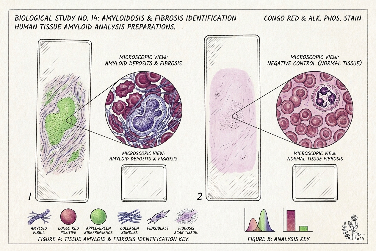
The detection toolkit here is consistent across labs. Thioflavin T binds the beta-sheet core of amyloid and fluoresces. Congo red gives apple-green birefringence under polarized light. Electron microscopy shows fibril shape. ThT kinetics track how quickly fibrils assemble.
That same ThT principle is what fibrin amyloid microscopy applies to plasma. Our pillar guide to amyloid fibril testing and ThT detection explains those methods, while the amyloid fibrin test guide focuses on microclots in blood. To see how commercial antibody and antigen panels compare with downstream clot detection, see our guide on spike protein test options and microclot analysis. If you're working out which test answers which question, the comparison of types of amyloid tests lines up Alzheimer's blood tests, PET, CSF, amyloidosis biopsy and fibrin amyloid microscopy.
Where our test fits
Our at-home microclot test is a finger-prick dried blood sample mailed to a partner laboratory in Mexico City. The lab stains it with Thioflavin T and records 6K 12-bit RAW video microscopy at 20x magnification under blue light, reviewed with AI-assisted fibril detection plus trained human oversight. The result is a Videographic Microscopy Report in a secure portal.
It's laboratory observational data for informational and research purposes, not a diagnosis and not a medical device. It doesn't measure circulating spike protein, which needs a separate spike-specific assay, and it doesn't quantify D-dimer or fibrinogen. Our comparison of D-dimer versus microclot testing explains why a normal D-dimer doesn't settle the microclot question.
What to read next
If clotting is your main concern, the amyloid fibrin test guide goes deeper, and the research on nattokinase and lumbrokinase for microclots covers what's been studied for breaking fibrinaloids down, while microclot treatment options sets out every approach and its evidence. Bring any result to a clinician who can read it alongside your history and standard labs.
Frequently asked questions
Do spike protein amyloid fibrils cause Long COVID?
That has not been shown. The 2025 study proposes a mechanism: Spike685 fibrils produce fibrin clots that resist breakdown, which could feed the fibrin amyloid microclots reported in Long COVID. The work used purified proteins in a test tube. Researchers still need to show that these fibrils form in people, at what levels, and whether they track with symptoms before anyone can call this a cause.
How are spike amyloid fibrils detected in the lab?
Several checks are used together. Thioflavin T is a dye that fluoresces when it binds the stacked beta-sheet core of amyloid. Congo red staining, electron microscopy and ThT assembly kinetics add confirmation. The 2022 paper from the same group required ThT kinetics, Congo red positivity and fibril morphology together before calling a spike peptide a genuine amyloid.
Does the vaccine spike protein contain the Spike685 sequence?
The original mRNA constructs encode full-length spike with two stabilizing proline changes, so residues 685 to 701 are part of the encoded protein. In their discussion, Westman and colleagues hypothesize that early constructs, which retained the furin cleavage site, could reach a cleaved aggregation-prone state. They present that as a hypothesis. No vaccinated patients were studied in the paper.
Can a standard blood test show fibrinolysis-resistant clots?
Usually not. Routine tests such as D-dimer and fibrinogen measure breakdown products or clotting proteins, not whether small amyloid-like clots are present. Research groups use ThT fluorescence microscopy on plasma to see fibrin amyloid microclots directly. Neither approach detects spike fibrils specifically, which would need a spike-specific assay.
Sources
- SARS-CoV-2 Spike Protein Amyloid Fibrils Impair Fibrin Formation and Fibrinolysis. Biochemistry, 2025
- Amyloidogenesis of SARS-CoV-2 Spike Protein. Journal of the American Chemical Society, 2022
- SARS-CoV-2 spike protein S1 induces fibrin(ogen) resistant to fibrinolysis: implications for microclot formation in COVID-19. Bioscience Reports, 2021
- A central role for amyloid fibrin microclots in long COVID/PASC: origins and therapeutic implications. Biochemical Journal, 2022
- Persistent clotting protein pathology in Long COVID/Post-Acute Sequelae of COVID-19 (PASC) is accompanied by increased levels of antiplasmin. Cardiovascular Diabetology, 2021
At-Home Amyloid Test
Curious whether fibrin amyloid microclots show up in your blood?
Our at-home amyloid fibrin test uses Thioflavin T fluorescence microscopy and returns a videographic report you can share with your doctor.
Next steps
- Spike Protein Test: Antibody, Antigen, and Downstream TestingLearn how spike protein tests work. Why antibody assays measure memory rather than active protein, and how downstream amyloid testing reveals microclots.
- Amyloid Fibril Test: Methods Used to Detect Amyloid FibrilsHow an amyloid fibril test works: Thioflavin T, Congo red, electron microscopy, X-ray diffraction and seeding assays compared, with which are clinical.
- What Are Amyloid Microclots? Definition, Causes and EvidenceWhat are amyloid microclots? A plain definition of these fibrin particles in blood samples, how they form, what studies found and what Cochrane says.

Tim Ottowitz
Founder & Research Lead
Tim Ottowitz founded Amyloid Test and writes its Science Hub, turning fibrin amyloid and spike protein research into plain-language guides that stay close to the published evidence.
Keep reading
Neutrophil Elastase and Spike Protein: How Amyloid Forms
Neutrophil elastase cuts SARS-CoV-2 spike into amyloid-forming fragments. A plain-English walkthrough of the 2022 JACS study and what it does not show.
Spike Protein Antibody vs Antigen Testing: What Each One Measures
Spike antibody tests measure immune memory, not spike protein. What antigen testing adds, why detox protocols stall, and what a serious panel includes.
Microclots and the COVID Vaccine: What the Spike-Fibrin Research Shows
Microclots and the COVID vaccine: what spike-fibrin lab studies and a 2026 cohort of 14 people show, and where the evidence stops today.