Neutrophil Elastase and Spike Protein: How Amyloid Forms
Short answer
Neutrophil elastase is an enzyme your white blood cells release during infection. In a 2022 study at Linköping University, it cut purified SARS-CoV-2 spike protein into fragments that formed amyloid fibrils, and clots made in the presence of those fibrils resisted breakdown. Intact folded spike didn't form fibrils on its own. All of this was done in test tubes, not in people.
Key takeaways
- 1A computational scan found seven amyloid-prone stretches in the spike protein, and three met every amyloid criterion when synthesized and tested.
- 2Full-length folded spike did not form amyloid fibrils by itself; it had to be cleaved first.
- 3Neutrophil elastase cleaved spike efficiently and released the amyloidogenic peptide 194 to 203.
- 4Fibrils formed from spike after elastase treatment were branched, which the authors noted is unusual for amyloid.
- 5Clots formed with spike amyloid present resisted plasmin, but the whole study used purified proteins in vitro.
If you've read that your own immune system can turn spike protein into amyloid, this is the paper behind that claim. It was published in the Journal of the American Chemical Society in May 2022 by two researchers at Linköping University in Sweden. Here's what they did, what they found, and the boundaries they drew around it.
What the 2022 study asked
Severe and long-lasting COVID-19 often comes with problems well beyond the lungs: disturbed coagulation, neurological trouble, cardiac complications. Sofie Nyström and Per Hammarström noticed those patterns resemble what's seen in diseases driven by misfolded amyloid proteins (Chiti & Dobson, 2017).
So they asked whether the spike protein itself contains amyloid-forming sequences, and if so, what would expose them (Nyström & Hammarström, 2022).
Start with the vocabulary
What is amyloid?
Amyloid is a folding pattern, not a single substance. When certain proteins misfold and stack against each other, they form a dense, highly stable structure the body struggles to break apart. Over 30 human proteins are known to adopt this disease-associated form (Chiti & Dobson, 2017).
Think of a shirt that gets twisted, damp, and then stuck to five other shirts. The pile is far harder to deal with than any single shirt.
What is neutrophil elastase?
Neutrophils are the white blood cells that arrive first and in large numbers during an infection. When they activate, they release neutrophil elastase, an enzyme that cuts proteins apart. It's one of the most abundant enzymes at inflamed sites of viral infection (Korkmaz et al., 2010).
Neutrophils do more than release enzymes. They also throw out webs of DNA called NETs, and our explainer on microclots, NETs and Long COVID covers how those webs tie into clotting, with DNase research on NET clearance looking at what might break them down.
What is the spike protein?
Spike is the protein on the surface of SARS-CoV-2 that lets the virus enter cells. It's 1,273 amino acids long and the part of the virus your immune system sees most clearly. It's also the protein that mRNA vaccines instruct cells to make.
What did the researchers actually do?
Their sequence of experiments was straightforward.
They predicted where amyloid might form. The WALTZ algorithm scans an amino acid chain and flags regions likely to misfold into amyloid (Maurer-Stroh et al., 2010). It found seven candidate regions in spike.
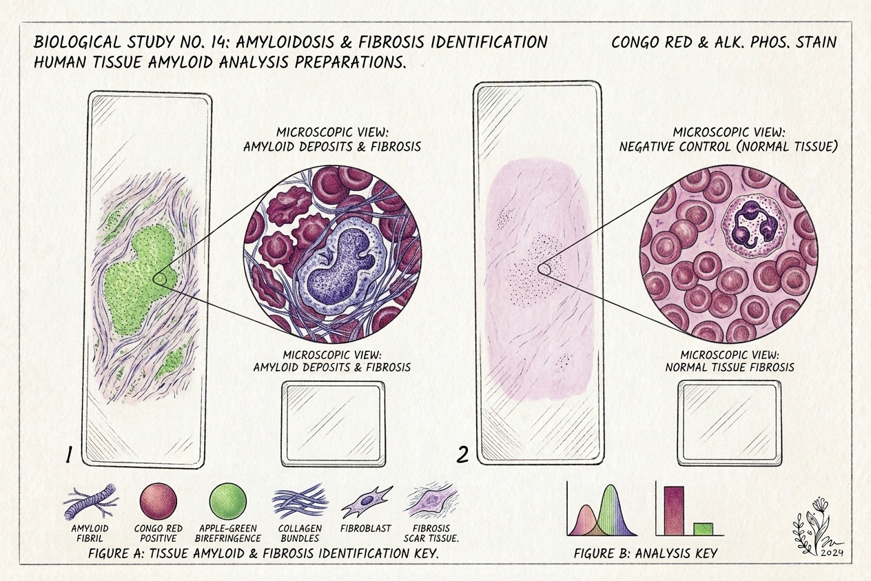
They synthesized those regions and tested them. Each predicted sequence was made as a short peptide and run through three standard amyloid checks: Thioflavin T fluorescence, Congo red birefringence under polarized light, and electron microscopy for fibril shape.
They mixed full-length spike with neutrophil elastase. Then they analyzed which fragments came out.
They tested the effect on clotting. Spike amyloid was combined with fibrinogen and thrombin, a clot was formed, and plasmin was added to see whether the clot dissolved normally.
What they found
Seven amyloid-prone regions, three confirmed
All seven predicted peptides aggregated when incubated at 37 degrees Celsius. Three met all three amyloid criteria: the peptides spanning residues 192 to 211, 601 to 620 and 1166 to 1185. The rest aggregated without meeting the full definition.
Not every suspicious-looking stretch of protein makes real amyloid, and that distinction matters when people summarize this work.
Intact spike did not form amyloid on its own
Tested by itself, full-length folded spike protein didn't form amyloid fibrils. Something had to cut it open first. That single result is what makes the next finding important.
Neutrophil elastase exposed the amyloidogenic segments
Elastase cleaved spike efficiently, and among the fragments it produced was peptide 194 to 203, part of the most amyloidogenic synthetic peptide the team had identified. In other words, the enzyme your own neutrophils release generated the kind of fragment that forms amyloid.
The fibrils were branched
After 24 hours of incubating spike with elastase, the fibrils under the electron microscope showed evident branching. Ordinary amyloid fibrils are straight and unbranched, so the authors called this out as an unusual morphology tied to the spike protein's own structure.
Clots with spike amyloid resisted breakdown
In the functional test, fibrinogen plus thrombin produced a normal clot that plasmin dissolved on schedule. With amyloid-forming spike fragments present, the resulting clot resisted plasmin. Turbidity measurements, the standard way of tracking clot formation and lysis, showed the difference.
A South African group had already reported that recombinant spike S1 could induce fibrin resistant to fibrinolysis in plasma (Grobbelaar et al., 2021). This paper offered a molecular reason why.
Clinical next step
Want to know whether amyloid shows up in your own blood?
Our at-home amyloid fibrin test stains a dried blood sample with Thioflavin T and returns a videographic microscopy report for you and your clinician.
Start screeningWhy does this matter for Long COVID?
The proposed chain runs: infection, neutrophil influx, elastase release, spike cleavage, amyloidogenic fragments, amyloid fibrils, clots that resist breakdown.
If that chain operates in people, it would speak to several puzzles. Standard blood work often looks normal in Long COVID while specialized fluorescence microscopy shows small amyloid-positive clots, a pattern documented in the fibrin amyloid microclot research. Amyloid structures are famously long-lived, which fits symptoms that persist. And amyloid can deposit in many tissues, which fits a multi-system illness. Our plain-language guide to amyloid microclots in Long COVID walks through the patient-level evidence.
A 2025 follow-up from the same group sharpened the picture considerably. Testing each of the seven spike amyloids against purified clotting components, they found that Spike601 and Spike685 behave in distinct ways while the other five did little (Westman et al., 2025).
What the paper does not show
It doesn't show this happens in patients. Every experiment was in a test tube with purified proteins. The abstract itself says the data propose a mechanism for potential amyloidogenesis in humans.
It doesn't prove spike amyloid causes Long COVID symptoms. A plausible biochemical mechanism isn't clinical evidence.
It doesn't establish how much spike is needed. The lab concentrations may differ substantially from anything that occurs in a person.
It doesn't cover variants, other coronaviruses, or vaccine-derived spike. The study used the original Wuhan-strain sequence. Whether similar processing happens after vaccination is a question the paper raises rather than answers, and our review of microclots reported after mRNA vaccination covers what the small cohorts have and haven't found since.
It doesn't mean every exposure leads here. Infection severity, the intensity of the neutrophil response, and individual differences in protein clearance would all matter.
How is amyloid detected in blood?
The same dyes used in this paper are what detection rests on. Thioflavin T fluoresces when it binds the beta-sheet core of amyloid, which is why it's the workhorse of both laboratory amyloid research and fibrin amyloid microscopy on plasma. Our pillar guide to amyloid fibril testing with Thioflavin T explains the method in detail, while the amyloid fibrin test guide focuses on microclots specifically.
If you're trying to figure out which test answers which question, the overview of types of amyloid tests compares Alzheimer's blood tests, PET, CSF testing, amyloidosis biopsy and fibrin amyloid microscopy. And because people often ask why their D-dimer came back fine, there's a separate comparison of D-dimer versus microclot testing.
Our own at-home amyloid test applies the Thioflavin T staining principle to a finger-prick dried blood sample, with 6K video microscopy reviewed by AI-assisted detection and trained human oversight. It's laboratory observational data, not a diagnosis, and it doesn't measure spike protein itself.
The bottom line
This isn't a clinical trial and it won't produce a treatment tomorrow. What it does is identify a specific, testable pathway by which a viral protein, processed by the host's own enzymes, can become a disease-associated form that disrupts normal clotting.
The mechanism is plausible and the experiments are clean. The jump from "this happens in a test tube" to "this drives symptoms in patients" hasn't been made. Hold both of those at once, and talk through anything you read here with a clinician who knows your history.
Frequently asked questions
What does neutrophil elastase do in the body?
Neutrophils are first-responder white blood cells. When they activate, they release neutrophil elastase, an enzyme that cuts proteins apart as part of killing pathogens and remodelling tissue. It's abundant at inflamed sites of viral infection. In COVID-19, neutrophils are recruited heavily to the lungs and other tissues, which is why the enzyme became the focus of this study.
Did the study prove spike protein turns into amyloid in patients?
No. Every experiment used purified proteins in a test tube. The authors' own abstract says the data propose a molecular mechanism for potential amyloidogenesis in humans. Whether this cleavage and fibril formation happens in living people at meaningful levels, and whether it contributes to symptoms, is still unknown.
Why were the spike fibrils described as branched?
Amyloid fibrils are usually long, straight and unbranched. When the researchers incubated full-length spike with neutrophil elastase for 24 hours and looked under the electron microscope, they saw amyloid-like fibrils with evident branching. The authors flagged this as an unusual morphology and linked it to the structure of the spike protein itself.
Which spike sequences formed real amyloid?
Of the seven predicted sequences, three met all three criteria the team set: nucleation-dependent assembly measured by Thioflavin T, Congo red positivity, and fibrillar shape under electron microscopy. Those were the 20-amino-acid peptides spanning residues 192 to 211, 601 to 620 and 1166 to 1185. The remaining four aggregated but did not meet the full definition.
Sources
- Amyloidogenesis of SARS-CoV-2 Spike Protein. Journal of the American Chemical Society, 2022
- SARS-CoV-2 Spike Protein Amyloid Fibrils Impair Fibrin Formation and Fibrinolysis. Biochemistry, 2025
- Protein Misfolding, Amyloid Formation, and Human Disease. Annual Review of Biochemistry, 2017
- Exploring the sequence determinants of amyloid structure using position-specific scoring matrices. Nature Methods, 2010
- Neutrophil Elastase, Proteinase 3, and Cathepsin G as Therapeutic Targets in Human Diseases. Pharmacological Reviews, 2010
- SARS-CoV-2 spike protein S1 induces fibrin(ogen) resistant to fibrinolysis. Bioscience Reports, 2021
At-Home Amyloid Test
Want to know whether amyloid shows up in your own blood?
Our at-home amyloid fibrin test stains a dried blood sample with Thioflavin T and returns a videographic microscopy report for you and your clinician.
Next steps
- Spike Protein Amyloid Fibrils: How Spike685 Makes Clots Resist LysisHow spike protein amyloid fibrils change fibrin clots: what the 2025 Linköping study found for Spike601 and Spike685, and what it cannot yet tell us.
- Amyloid Fibril Test: Methods Used to Detect Amyloid FibrilsHow an amyloid fibril test works: Thioflavin T, Congo red, electron microscopy, X-ray diffraction and seeding assays compared, with which are clinical.
- Neutrophil Extracellular Traps in Long COVID: The Microclot-NET StudyA 2025 study found neutrophil extracellular traps tangled into microclots in Long COVID. What the paper measured, what it found, and what it can't prove.

Tim Ottowitz
Founder & Research Lead
Tim Ottowitz translates peer-reviewed research on spike protein pathology, amyloid biology and post-viral syndromes into clear, carefully sourced patient education.
Keep reading
Spike Protein Antibody vs Antigen Testing: What Each One Measures
Spike antibody tests measure immune memory, not spike protein. What antigen testing adds, why detox protocols stall, and what a serious panel includes.
SARS-CoV-2 Cell-to-Cell Spread: Nanotubes, Syncytia and Vesicles
How might SARS-CoV-2 spread from cell to cell without leaving? A simple guide to the nanotubes, fused cells and vesicles seen in a 2022 microscopy study.
COVID Vaccine Harms: What the 2025 JPandS Review Argues
A plain-language walkthrough of the 2025 JPandS review on COVID vaccine harms: what it claims, which sources hold up, and where the evidence is weak.