Lipid Nanoparticle Blood Distribution: How mRNA Circulates in Humans
Short answer
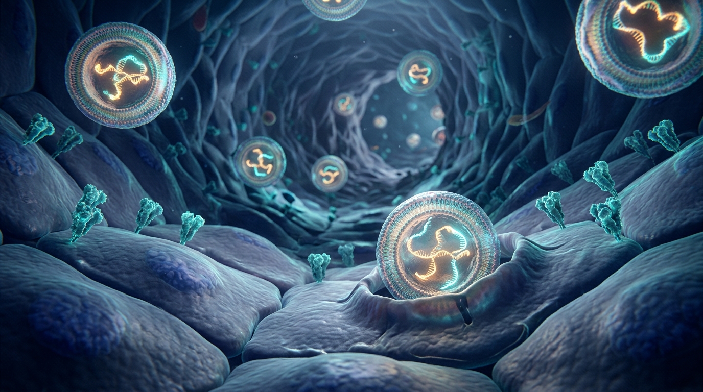
A 2024 human pharmacokinetic study in ACS Nano demonstrated that lipid nanoparticles injected into arm muscle enter the bloodstream, with vaccine mRNA and the ionizable lipid SM-102 peaking at 1 to 2 days post-vaccination. Key takeaway: Both components cleared in parallel and remained quantifiable in 37 percent of human participants up to 15 days, proving that intact nanocarriers circulate systemically.
Key takeaways
- 1Serial blood testing in vaccinated humans showed peak systemic blood concentrations of mRNA (0.19 ng/mL) and SM-102 lipid (3.22 ng/mL) on days 1 to 2.
- 2Identical decay rates between mRNA and the ionizable lipid indicate that lipid nanoparticles circulate as intact complexes rather than degraded fragments.
- 3Vaccine mRNA remained detectable in the bloodstream of 37 percent of human subjects out to days 14 and 15 following booster vaccination.
- 4Monocyte phagocytosis serves as a primary clearance mechanism, while anti-PEG antibodies interact with the outer lipid shell without completely blocking circulation.
- 5Circulating nanocarriers deliver genetic cargo to capillary endothelial cells, offering a direct mechanism for microvascular inflammation and downstream microclots.
Early public health guidance stated that mRNA vaccines remained confined to the muscle tissue of the upper arm. A landmark 2024 clinical study published in ACS Nano by Stephen Kent and colleagues challenged this assumption by tracking serial blood samples in vaccinated humans. In this article, you will learn how the study measured circulating mRNA and ionizable lipids in human blood, how these particles interact with immune cells, and how their systemic transit connects to microvascular inflammation and microclot formation.
Understanding lipid nanoparticle blood distribution bridges the gap between local intramuscular injection and the systemic vascular symptoms reported by patients with post-vaccination syndrome.
Human Pharmacokinetics: Tracking mRNA and SM-102 in the Bloodstream
Previous biodistribution evidence relied largely on preclinical animal models. The 2024 ACS Nano investigation provided the first comprehensive human pharmacokinetic profile of both primary components of the Moderna SPIKEVAX mRNA booster: the genetic mRNA payload and the proprietary ionizable lipid carrier, SM-102.
Researchers collected serial venous blood draws from human volunteers at regular intervals, ranging from hours after vaccination up to 28 days post-injection. Using ultra-sensitive reverse transcription quantitative PCR (RT-qPCR) for the vaccine mRNA and high-performance liquid chromatography tandem mass spectrometry (LC-MS/MS) for the SM-102 lipid, the team mapped exact circulation kinetics.
The study established several critical findings:
- Rapid Blood Entry: Both vaccine mRNA and SM-102 lipids escaped the deltoid muscle and entered systemic circulation within hours of administration.
- Peak Timing: Both components reached peak systemic concentrations between 24 and 48 hours (days 1 to 2) post-vaccination.
- Quantified Peak Levels: Median peak blood concentrations reached approximately 0.19 nanograms per milliliter (ng/mL) for intact mRNA and 3.22 ng/mL for the ionizable lipid SM-102.
- Synchronized Clearance: The elimination slopes of mRNA and SM-102 mirrored each other closely. This parallel decay proves that lipid nanoparticles circulate in human blood as intact, assembled nanocarrier spheres rather than breaking apart immediately into free components at the injection site.
- Extended Circulation Window: While blood levels fell significantly by day 7, quantifiable levels of vaccine mRNA remained detectable in the bloodstream of 37 percent of participants out to days 14 and 15.
These findings directly corroborate earlier observational work by Ogata and colleagues, who detected circulating spike antigen in human plasma following vaccination, and confirm that the vascular system experiences direct exposure to intact carrier particles.
Clearance Biology: Monocytes and Anti-PEG Antibodies
Lipid nanoparticles rely on a four-component lipid formulation. In addition to cholesterol, helper phospholipids, and the ionizable lipid SM-102, the outer surface is coated with PEGylated lipids (PEG2000-DMG). Polyethylene glycol (PEG) forms a hydrophilic protective brush that prevents particles from clumping and delays clearance by the liver's reticuloendothelial system.
The ACS Nano study investigated how the immune system interacts with this synthetic PEG shell in human blood:
Monocyte Phagocytosis
The authors showed that circulating monocytes, a type of white blood cell, are actively involved in engulfing circulating lipid nanoparticles from human blood. Monocytes internalize the particles via endocytosis, transporting the foreign lipid and RNA cargo through the bloodstream toward the spleen and liver for metabolic degradation.
Anti-PEG Antibody Dynamics
Because PEG is widely used in cosmetics, pharmaceuticals, and consumer products, many individuals possess pre-existing anti-PEG antibodies. The study evaluated whether pre-existing antibodies to PEG accelerated the clearance of circulating nanocarriers.
While pre-existing anti-PEG IgG and IgM titers varied widely across participants, they did not prevent lipid nanoparticles from circulating during the initial 48-hour window. However, systemic exposure to the vaccine particles triggered a significant boost in anti-PEG antibody levels over subsequent weeks. This antibody surge demonstrates that circulating nanocarriers interact directly with the systemic humoral immune response.
Preclinical Models vs. Human Clinical Data
The human blood distribution data from ACS Nano connects directly to the tissue-level mechanisms uncovered in recent preclinical imaging studies.
The table below summarizes how human clinical pharmacokinetics compare with high-resolution animal imaging and downstream vascular assessments:
| Investigation Level | Model & Technique | Key Biological Observation | Clinical Significance |
|---|---|---|---|
| Whole-Body Biodistribution | Mouse models via optical clearing and SCP-Nano 3D imaging (Nature Biotech 2025) | Injected LNPs escape muscle and localize heavily to liver, spleen, and capillary endothelial cells in heart tissue. | Proves that distant microvascular beds directly uptake circulating lipid nanoparticles. |
| Human Blood Pharmacokinetics | Human serial blood draws via RT-qPCR and LC-MS/MS (ACS Nano 2024) | Intact mRNA and SM-102 peak in human blood at days 1–2, persisting up to 15 days in 37% of subjects. | Confirms systemic circulation of intact nanocarriers in living human patients. |
| Cellular Membrane Stress | In vitro and animal lipid bilayer studies (IJMS 2023) | Ionizable cationic lipids disrupt cell membrane fluidity, alter calcium signaling, and trigger intracellular stress. | Explains non-specific inflammatory signaling and endothelial activation. |
| Downstream Vascular Pathology | Thioflavin T fluorescence microscopy of human blood (Biochemical Journal 2022) | Misfolded fibrin forms plasmin-resistant amyloid microclots that entrap platelets and occlude microcapillaries. | Provides an objective physical readout of ongoing microvascular hypoperfusion. |
From Circulating Nanocarriers to Endothelial Microclots
Why does the systemic circulation of lipid nanoparticles matter for patient symptoms? The answer lies in the interaction between circulating nanocarriers and the microvasculature.
As detailed in our review of how two blood drops detect fibrils, blood completes a full circulatory transit through the heart and body roughly once every 60 seconds. When intact lipid nanoparticles circulate continuously through human blood for days or weeks, they make repeated contact with endothelial cells lining microscopic capillaries:
- Endothelial Uptake: Capillaries have narrow lumens measuring only 5 to 10 micrometers across. Circulating nanocarriers slow down in these peripheral beds, allowing endothelial cells to internalize the particles via endocytosis.
- Spike Synthesis in Blood Vessels: Once inside endothelial cells, the mRNA instructs the cell's ribosomes to manufacture SARS-CoV-2 spike protein. The cell displays this foreign antigen on its luminal surface facing the bloodstream.
- Endothelial Activation and Shear Stress: Surface expression of spike protein triggers local immune recognition, downregulates protective endothelial surface markers like CD34, and induces vascular shear stress.
- Amyloid Fibrin Formation: Circulating soluble fibrinogen directly contacts membrane-bound and shed spike protein. As documented in research on post-vaccination microclots, spike protein induces a conformational transition in fibrinogen, forcing it to polymerize into anomalous, beta-sheet-rich fibrin amyloid microclots.
- Microvascular Occlusion: Because these anomalous microclots resist breakdown by plasmin, they remain lodged in peripheral microcapillaries, reducing oxygen delivery and driving chronic fatigue, brain fog, and exercise intolerance.
Standard clinical coagulation panels, such as D-dimer, fail to capture this process because they only measure fragments generated during normal clot breakdown, leaving patients in a diagnostic void.
Where Our At-Home Amyloid Test Fits
For patients experiencing persistent symptoms after viral exposure or vaccination, knowing whether microvascular pathology is present is an essential first step. Standard commercial labs do not offer assays to track circulating lipid nanoparticles or measure nanogram-level SM-102.
Our at-home amyloid test addresses this gap by assessing the downstream microvascular consequences of vascular inflammation:
- At-home dried blood spot: You collect a small capillary blood sample using a gentle lancet and mail it to our partner laboratory in Mexico City.
- Thioflavin T (ThT) staining: Technicians stain the specimen with Thioflavin T, an optical dye that binds specifically to cross-beta sheet amyloid fibrils.
- 6K 12-bit RAW video microscopy: The lab records multi-field digital video at 20x magnification under ultraviolet illumination.
- AI-assisted detection with human review: Computer-vision algorithms quantify microclot burden, verified by trained analysts.
- Objective visual report: You receive a detailed Videographic Microscopy Report in a secure portal within 1 day of laboratory arrival (typically 5 to 7 business days from ordering).
- Transparent pricing: The test costs $160 USD (3,200 MXN), avoiding the multi-thousand-dollar cash fees charged by private research clinics.
What Our Test Does Not Do
To maintain scientific integrity and adhere to regulatory guidelines:
- Not a diagnostic test or medical device: Our test provides observational laboratory data for research, wellness, and informational tracking. It does not replace medical consultation.
- Does not detect lipid nanoparticles or measure SM-102: It evaluates the downstream structural formation of fibrin amyloid microclots.
- Does not quantify circulating spike protein: While spike protein initiates microclot formation, direct antigen measurement requires separate specialized assays as discussed in our spike protein test overview.
- Not an Alzheimer's or systemic organ amyloidosis test: It does not evaluate brain amyloid PET plaques, CSF peptides, or organ-bound AL/ATTR amyloidosis.
To understand how testing methodologies compare, read our breakdown of at-home microclot tests vs clinic testing and our review of flow clotometry vs microscopy.
Next Steps for Patients and Clinicians
The 2024 ACS Nano study by Kent and colleagues conclusively showed that lipid nanoparticles and their mRNA cargo enter the systemic bloodstream in humans, peaking at 1 to 2 days and remaining detectable for over two weeks in a significant percentage of recipients.
If you are navigating persistent post-vaccination or post-viral symptoms, tracking microvascular health provides actionable objective context. Learn more by exploring our comprehensive guide on what microclots are and examining the clinical evidence behind the amyloid fibrin test.
Frequently asked questions
Do lipid nanoparticles enter the bloodstream after an intramuscular injection?
Yes. Serial blood sampling in human volunteers published in ACS Nano proved that lipid nanoparticles leave the deltoid muscle and enter systemic circulation. Both the mRNA genetic payload and the ionizable lipid SM-102 reached peak blood levels 1 to 2 days post-vaccination before gradual clearance.
How long do lipid nanoparticles circulate in human blood?
In the 2024 ACS Nano study, lipid nanoparticles cleared from blood over several days in most participants, but intact vaccine mRNA remained detectable and quantifiable in 37 percent of recipients up to 14 to 15 days post-injection. Clearance is largely mediated by circulating monocyte phagocytosis.
Do lipid nanoparticles circulate as intact particles or broken fragments?
They circulate largely as intact nanocarrier complexes. Researchers observed that the clearance kinetics of the mRNA core and the SM-102 lipid shell followed virtually identical decay slopes over two weeks, which would not happen if the lipid shell dissolved immediately at the injection site.
What are anti-PEG antibodies and how do they affect clearance?
Polyethylene glycol (PEG) coats the outer surface of lipid nanoparticles to prevent premature immune destruction. Exposure boosts anti-PEG antibodies in blood. The ACS Nano trial found that while baseline PEG antibodies did not halt circulation, post-vaccine antibody surges influenced subsequent clearance pathways.
Can standard blood tests detect circulating lipid nanoparticles?
No. Standard clinical laboratories lack assays to detect lipid nanoparticles or measure nanogram-level SM-102. Specialist research uses quantitative RT-PCR for mRNA and liquid chromatography mass spectrometry for lipids. Functional testing instead focuses on downstream vascular consequences, such as microvascular microclots.
Sources
- Blood Distribution of SARS-CoV-2 Lipid Nanoparticle mRNA Vaccine in Humans. ACS Nano, 2024
- Nanocarrier imaging at single-cell resolution across entire mouse bodies with deep learning. Nature Biotechnology, 2025
- Lipid Nanoparticles for Delivery of Messenger RNA to the Skin Induce Systemic Membrane Dysfunction. International Journal of Molecular Sciences, 2023
- Circulating Severe Acute Respiratory Syndrome Coronavirus 2 (SARS-CoV-2) Vaccine Antigen Detected in the Plasma of mRNA-1273 Vaccine Recipients. Clinical Infectious Diseases, 2021
- A central role for amyloid fibrin microclots in long COVID/PASC: origins and therapeutic implications. Biochemical Journal, 2022
At-Home Amyloid Test
Concerned about circulating spike, lipid carriers, or vascular symptoms?
Explore our at-home amyloid test kit to check for downstream fibrin amyloid microclots.
Next steps
- Spike Protein Amyloid Fibrils: How Spike685 Makes Clots Resist LysisHow spike protein amyloid fibrils change fibrin clots: what the 2025 Linköping study found for Spike601 and Spike685, and what it cannot yet tell us.
- Amyloid Fibril Test: Methods Used to Detect Amyloid FibrilsHow an amyloid fibril test works: Thioflavin T, Congo red, electron microscopy, X-ray diffraction and seeding assays compared, with which are clinical.
- Post-Vaccination Syndrome: Symptoms, Duration and the Spike ResearchPost-vaccination syndrome symptoms, how long it can last, and what spike persistence, spike amyloid fibrils and microclot studies show about the cause.

Tim Ottowitz
Founder & Research Lead
Tim Ottowitz founded Amyloid Test and writes its Science Hub, turning fibrin amyloid and spike protein research into plain-language guides that stay close to the published evidence.
Keep reading
Lipid Nanoparticle Biodistribution: Where mRNA Goes in the Body
A plain-English guide to lipid nanoparticle biodistribution research, showing how whole-body imaging tracks mRNA delivery to heart capillaries.
Lipid Nanoparticle Membrane Dysfunction: What the Omics Data Show
A plain-English guide to lipid nanoparticle membrane dysfunction, explaining how ionizable lipids alter cell signaling, metabolism, and membranes.
Spike Protein Antibody vs Antigen Testing: What Each One Measures
Spike antibody tests measure immune memory, not spike protein. What antigen testing adds, why detox protocols stall, and what a serious panel includes.