Lipid Nanoparticle Membrane Dysfunction: What the Omics Data Show
Short answer
Lipid nanoparticle membrane dysfunction describes how the fatty carriers used in mRNA medicines interact directly with cell membranes. A comprehensive 2026 review in Acta Pharmaceutica Sinica B analyzed transcriptomic and proteomic data, showing that ionizable lipids disrupt the phosphatidylinositol cycle, alter cell membrane curvature, and trigger stress cascades including NF-κB and MAPK, even without mRNA cargo present.
Key takeaways
- 1Lipid nanoparticles are not biologically inert envelopes; their ionizable lipids actively interact with cellular membranes.
- 2A 2026 review introduced the concept of lipid-nanoparticle-driven membrane dysfunction (L-DMD) based on multi-omics data.
- 3Empty lipid nanoparticles without any mRNA cargo alter gene expression, downregulate cytochrome P450 enzymes, and activate inflammatory signaling.
- 4The primary disruption occurs in the phosphatidylinositol cycle, which governs membrane curvature, endocytosis, and organelle repair.
- 5These biophysical membrane effects provide a systems-level framework for understanding prolonged cellular stress after mRNA exposure.
When public health agencies and pharmaceutical developers describe mRNA vaccines, the lipid delivery carrier is often treated as a passive wrapper. The conventional assumption has been that the fat bubble simply protects the fragile genetic sequence, drops it into the cell cytoplasm, and quietly dissolves without lasting consequences.
A comprehensive 2026 review published in Acta Pharmaceutica Sinica B by Seger, Gutschi, and Seneff challenges that passive model directly (Seger et al., 2026). Synthesizing transcriptomic, proteomic, and biophysical data, the authors explain that lipid nanoparticles (LNPs) act as biologically active interfaces that alter membrane mechanics, disrupt vital lipid signaling cycles, and initiate sustained intracellular stress.
By the end of this guide, you will understand what lipid nanoparticle membrane dysfunction means, how ionizable lipids interact with cell membranes, and what the omics data show about downstream cellular health.
What is lipid-nanoparticle-driven membrane dysfunction?
Living cell membranes are not static plastic sheets. They are dynamic, liquid-crystal bilayers made of phospholipids, cholesterol, and signaling lipids that maintain delicate electrical charges and distinct inner and outer leaflets.
Synthetic lipid nanoparticles rely on four main ingredients:
- Ionizable cationic lipids: Engineered to stay neutral in the bloodstream, but become positively charged inside acidic cellular compartments (endosomes) to disrupt the membrane and let mRNA escape.
- Cholesterol: Provides structural stability and supports membrane fusion.
- Helper phospholipids: Form the nanoparticle's spherical outer shell.
- PEGylated lipids: Polyethylene glycol coatings that shield the particle from immediate immune destruction.
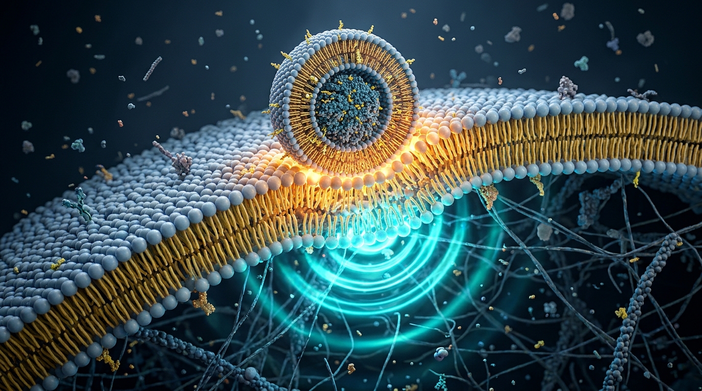
The review introduces the concept of lipid-nanoparticle-driven membrane dysfunction (L-DMD). The authors show that when ionizable lipids enter a cell, their cone-like geometry and positive charges insert directly into host membranes.
This physical integration disrupts normal membrane curvature, alters lipid distribution, and interferes with the cell's internal communication networks. Crucially, these effects occur whether the nanoparticle carries mRNA or is completely empty.
Because of these intrinsic membrane effects, researchers have increasingly investigated alternative carriers, such as protein nanoparticles for mRNA delivery, which avoid synthetic ionizable lipids entirely.
How do empty lipid nanoparticles affect cells?
One of the most persistent assumptions in pharmaceutical delivery is that delivery vehicles are biologically inert. But omics research (transcriptomics measuring RNA expression and proteomics measuring protein levels) shows that empty lipid nanoparticles (eLNPs) trigger widespread cellular responses on their own.
When cells or laboratory animals are exposed to empty lipid nanoparticles without any mRNA or antigen:
- Toll-like receptor 4 (TLR4) activation: The foreign lipid structures trigger cell surface receptors normally reserved for bacterial endotoxins, driving acute inflammatory signaling.
- Inflammatory cytokine surges: Transcriptomic studies consistently report elevations in interleukin-6 (IL-6), interferons, and chemokines like CXCL9 and CXCL10.
- Suppression of detoxification enzymes: Hepatic studies show marked downregulation of cytochrome P450 (CYP) enzymes, which are responsible for metabolizing medications and environmental toxins.
- Oxidative and mitochondrial stress: Membrane disruption increases reactive oxygen species (ROS), impairing cellular energy balance.
These findings mirror whole-body imaging studies on lipid nanoparticle biodistribution, which showed that empty nanoparticles alone alter hundreds of metabolic and translation proteins in off-target organs.
Next step
Concerned about lipid nanoparticles, spike persistence, or post-vaccine symptoms?
Explore screening options and discuss objective findings with a knowledgeable clinician.
See the at-home testWhy is the phosphatidylinositol cycle the central target?
To understand why membrane disturbance causes widespread problems, you have to look at the phosphatidylinositol (PI) cycle.
Phosphoinositides (PIPs) are specialized lipid molecules found on the inner surfaces of cellular membranes. Although they make up only a tiny fraction of membrane lipids, they act like biochemical ZIP codes. They tell the cell where to transport vesicles, how to repair damaged membranes, and when to activate growth or survival signals.
The 2026 review identifies the PI cycle as the primary vulnerability point where lipid nanoparticles disrupt cellular order:
1. Impaired endosomal repair and the ESCRT pathway
When an ionizable lipid breaches an endosome to release mRNA, it creates a physical hole. Cells rely on a molecular repair crew called the ESCRT complex (Endosomal Sorting Complex Required for Transport) to patch small tears before the compartment bursts.
ESCRT proteins depend directly on specific phosphoinositides to know where to assemble. Proteomic data show that LNP exposure downregulates ESCRT components and suppresses endocytic activity. When small tears cannot be repaired efficiently, endosomes and lysosomes rupture, releasing acidic contents and triggering the NLRP3 inflammasome.
2. Disrupted lipid metabolism and PPAR signaling
The review highlights that LNP exposure perturbs peroxisome proliferator-activated receptor gamma (PPARγ) pathways. Cells struggle to balance fatty acid oxidation, resulting in abnormal lipid droplet accumulation and metabolic exhaustion.
3. Altered membrane-bound drug metabolism
Cytochrome P450 enzymes sit embedded within the membranes of the endoplasmic reticulum and mitochondria. Their enzymatic activity relies on precise membrane fluidity and surrounding phospholipid geometry. When synthetic ionizable lipids alter the membrane environment, CYP enzymatic activity drops significantly, potentially altering how the body handles common drugs.
Comparison: Passive carrier model vs. L-DMD omics findings
The table below contrasts the traditional pharmacological view of lipid nanoparticles with the mechanisms documented in multi-omics literature.
| Cellular Process | Conventional Passive View | L-DMD Omics Evidence | Clinical & Biological Significance |
|---|---|---|---|
| Membrane Interaction | Assumed transient and biologically neutral after payload release | Ionizable lipids integrate into bilayers, altering curvature, charge, and leaflet symmetry | Disrupts membrane-embedded receptors and cellular mechanics. Discussed in post-vaccination microclots. |
| Empty Particle Bioactivity | Treated as an inert control vehicle | Drives TLR4 activation, NF-κB inflammatory cascades, and oxidative stress | Proves that the lipid formulation itself is intrinsically immunogenic. |
| Phosphoinositide (PI) Signaling | Ignored as unaffected background biochemistry | Perturbs PIP conversion and downregulates ESCRT membrane repair complexes | Leads to lysosomal leakage, cell stress, and NLRP3 inflammasome activation. |
| Drug Metabolism (CYP Enzymes) | Assumed unaffected by lipid delivery vehicles | Significant downregulation of cytochrome P450 enzymes across transcriptomes | May explain adverse drug interactions and altered medication clearance post-exposure. |
| Organelle Trafficking | Standard endocytosis assumed to proceed normally | Impairs vesicle recycling, receptor internalization, and mitochondrial respiration | Promotes prolonged cellular exhaustion and dysregulated signaling. Covered in spike protein persistence testing. |
How does membrane dysfunction amplify spike protein pathology?
When lipid nanoparticles deliver modified mRNA (modRNA) encoding the SARS-CoV-2 spike protein, membrane dysfunction does not occur in a vacuum. It interacts synergistically with the viral antigen being produced.
- Membrane stress meets viral protein: While ionizable lipids disrupt endosomal membranes and the PI cycle, the cell's ribosomes begin translating spike protein. Spike proteins are transmembrane structures that anchor directly into host cell membranes before shedding.
- Capillary endothelial vulnerability: As shown in biodistribution imaging, lipid nanoparticles reach the endothelial cells lining microscopic capillaries. When capillary lining cells suffer simultaneous membrane disruption and spike protein expression, local vascular stress escalates.
- Fibrinogen and microclot formation: Injured endothelial cells and circulating spike fragments promote abnormal blood clotting. Spike protein can interact directly with fibrinogen to form dense, breakdown-resistant microclots. To learn more about this mechanism, explore our guide on what are microclots.
By viewing lipid nanoparticles as active biointerfaces rather than passive carriers, researchers can better explain why a subset of individuals experiences prolonged fatigue, cardiovascular symptoms, or neurovascular complications following mRNA-LNP exposure.
What are the limitations of the current evidence?
While the L-DMD framework synthesizes hundreds of published studies, several important caveats apply:
- Mechanistic framework, not a standalone diagnostic test: The review integrates transcriptomic and proteomic data from cell cultures, animal models, and human observational cohorts. It provides a molecular explanation for observed phenomena, but does not provide a single diagnostic blood test for L-DMD.
- Variable resolution kinetics: The authors note that the exact time it takes for different cell types to fully clear ionizable lipids and restore the PI cycle remains uncertain and varies by dose, tissue, and individual genetics.
- Preclinical models dominate: Much of the detailed mechanistic work on membrane curvature, ESCRT downregulation, and organelle rupture comes from in vitro cell models and rodent experiments. Translating these findings to long-term human clinical outcomes requires dedicated prospective clinical trials.
Where does amyloid microclot testing fit?
When cellular membranes and vascular linings experience prolonged stress, the surrounding microvascular environment can shift toward chronic, low-grade clotting. In particular, spike protein exposure can drive fibrin to misfold into an amyloid-like state that resists normal enzymatic breakdown.
If you are dealing with unexplained post-viral or post-vaccine health complications, evaluating your blood for microvascular clotting provides objective, measurable information.
Our at-home amyloid test evaluates a dried blood spot collected via a simple finger prick. Technicians at our partner laboratory stain the sample with Thioflavin T (ThT) fluorescent dye and record high-resolution 6K video microscopy under UV light. AI-assisted fibril detection and trained human microscopists analyze the footage to observe amyloid-positive microclots.
It is important to understand the boundaries of the test:
- It does detect whether your blood contains fibrin amyloid microclots that resist normal fibrinolysis.
- It does not measure intracellular membrane disruption, lipid nanoparticle remnants, or circulating spike protein levels.
- It is not an Alzheimer's test, is not a test for systemic amyloidosis, and does not diagnose any illness.
You receive an objective Videographic Microscopy Report that you can review with your healthcare provider. For a detailed breakdown of how ThT fluorescence detects abnormal clotting, see our pillar guide on the amyloid fibrin test.
What should you do next?
Understanding the biophysical reality of lipid nanoparticles helps move the conversation beyond oversimplified assumptions. If you suspect you are dealing with lingering effects from viral exposure or lipid nanoparticle medicines, consider these constructive steps:
- Consult an informed healthcare provider: Share peer-reviewed literature detailing membrane dynamics, vascular health, and inflammatory signaling.
- Investigate microvascular clotting: Learn how persistent spike protein drives abnormal clot resistance in our guide on post-vaccination microclots.
- Explore antigen and persistence data: Review what current laboratory tests can and cannot detect in our breakdown of spike protein persistence testing.
This article is educational and does not provide medical advice, diagnosis, or treatment recommendations.
Frequently asked questions
What is lipid-nanoparticle-driven membrane dysfunction (L-DMD)?
Lipid-nanoparticle-driven membrane dysfunction (L-DMD) is a mechanistic framework describing how synthetic lipid nanoparticles disturb living cell membranes. When ionizable lipids fuse with or cross cellular and endosomal membranes, they alter membrane charge, curvature, and lipid symmetry. This physical disturbance interrupts the phosphatidylinositol cycle, triggering downstream inflammation, metabolic stress, and altered cellular communication throughout the cell.
Do empty lipid nanoparticles cause cellular changes without mRNA?
Yes. Multiple transcriptomic and proteomic studies reviewed in the 2026 paper demonstrate that empty lipid nanoparticles (eLNPs) without any mRNA payload alter hundreds of genes and proteins. They trigger Toll-like receptor 4 (TLR4) activation, induce nuclear factor kappa B (NF-κB) inflammatory signaling, suppress cytochrome P450 detoxification enzymes, and cause oxidative stress in cell and animal models.
What is the phosphatidylinositol cycle and why does it matter?
The phosphatidylinositol (PI) cycle is a vital biochemical network that generates phosphoinositide (PIP) signaling lipids on cell membranes. These lipids act as cellular area codes that guide membrane repair, endocytosis, and protein transport. When ionizable lipids perturb the PI cycle, cells struggle to repair ruptured endosomes, transport cellular cargo, and maintain balanced metabolic signaling.
Which cellular signaling pathways are affected by lipid nanoparticles?
Omics datasets show that lipid nanoparticles perturb several primary signaling networks. These include the NF-κB and MAPK pathways, which drive inflammatory cytokine production, the JAK/STAT pathway, which regulates immune activation, the mTOR pathway, which controls cellular growth and energy balance, and PPARγ signaling, which directs lipid metabolism and mitochondrial function.
Does amyloid microclot testing detect lipid nanoparticle membrane damage?
No. The Amyloid Test kit evaluates whether a dried blood sample contains fibrin amyloid microclots using Thioflavin T fluorescence microscopy. It does not measure intracellular membrane dysfunction, does not track lipid nanoparticles, and is not a diagnostic test. It provides observational laboratory data regarding abnormal clotting in circulation.
Sources
- Lipid nanoparticles as active biointerfaces: From membrane interaction to systemic dysregulation. Acta Pharmaceutica Sinica B, 2026
- Nanocarrier imaging at single-cell resolution across entire mouse bodies with deep learning. Nature Biotechnology, 2025
- The mRNA-LNP platform's lipid nanoparticle component used in preclinical vaccine studies is highly inflammatory. iScience, 2021
- SARS-CoV-2 spike protein S1 induces fibrin(ogen) resistant to fibrinolysis. Biochemical Journal, 2021
At-Home Amyloid Test
Concerned about lipid nanoparticles, spike persistence, or post-vaccine symptoms?
Explore screening options and discuss objective findings with a knowledgeable clinician.
Next steps
- Spike Protein Amyloid Fibrils: How Spike685 Makes Clots Resist LysisHow spike protein amyloid fibrils change fibrin clots: what the 2025 Linköping study found for Spike601 and Spike685, and what it cannot yet tell us.
- Amyloid Fibril Test: Methods Used to Detect Amyloid FibrilsHow an amyloid fibril test works: Thioflavin T, Congo red, electron microscopy, X-ray diffraction and seeding assays compared, with which are clinical.
- Post-Vaccination Syndrome: Symptoms, Duration and the Spike ResearchPost-vaccination syndrome symptoms, how long it can last, and what spike persistence, spike amyloid fibrils and microclot studies show about the cause.

Tim Ottowitz
Founder & Research Lead
Tim Ottowitz founded Amyloid Test and writes its Science Hub, turning fibrin amyloid and spike protein research into plain-language guides that stay close to the published evidence.
Keep reading
Lipid Nanoparticle Biodistribution: Where mRNA Goes in the Body
A plain-English guide to lipid nanoparticle biodistribution research, showing how whole-body imaging tracks mRNA delivery to heart capillaries.

Spike Protein Antibody vs Antigen Testing: What Each One Measures
Spike antibody tests measure immune memory, not spike protein. What antigen testing adds, why detox protocols stall, and what a serious panel includes.
Spike Protein Test: Antibody, Antigen, and Downstream Testing
Learn how spike protein tests work. Why antibody assays measure memory rather than active protein, and how downstream amyloid testing reveals microclots.