Lipid Nanoparticle Biodistribution: Where mRNA Goes in the Body
Short answer
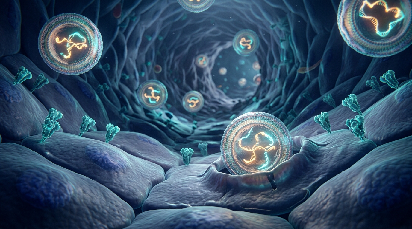
Lipid nanoparticle biodistribution studies track where tiny fat bubbles carrying mRNA travel after injection. A landmark 2025 Nature Biotechnology whole-body imaging study in mice revealed that intramuscularly injected lipid nanoparticles don't stay confined to the injection muscle. Instead, small amounts reach distant organs including the heart, where endothelial cells lining heart capillaries take them up, express spike protein, and show signs of vascular stress and immune activation.
Key takeaways
- 1A 2025 Nature Biotechnology study used whole-body 3D imaging in mice to track lipid nanoparticles at single-cell resolution.
- 2Intramuscularly injected lipid nanoparticles did not remain solely at the injection site, reaching distant organs including the liver, spleen, and heart.
- 3In the heart, mRNA-delivered spike protein localized specifically to endothelial cells lining capillaries, not heart muscle cells (cardiomyocytes).
- 4Heart capillary endothelial cells expressing spike protein showed measurable vascular stress markers and immune activation on mass spectrometry proteomics.
- 5The findings offer a clear biological mechanism for rare post-vaccine cardiac symptoms, though human clinical studies are still needed to confirm causation.
If you have ever received an mRNA shot, you might have been told that the contents stay right in your arm muscle. A 2025 study published in Nature Biotechnology used whole-body 3D imaging in mice to see where those tiny delivery packages actually travel. By the end of this guide, you will understand how researchers mapped lipid nanoparticles across entire bodies at single-cell resolution, what happened when spike mRNA reached the heart, and what these findings mean in simple, plain English.
What are lipid nanoparticles? (The delivery envelope)
Messenger RNA (mRNA) carries instructions that tell your cells how to make a specific protein. But naked mRNA is fragile. If you injected bare mRNA into muscle tissue, enzymes in your body would destroy it in minutes.
To protect the message, scientists pack mRNA inside tiny artificial bubbles made of fats, called lipid nanoparticles (LNPs). Think of an LNP as a protective delivery envelope. It shields the genetic letter during transit and fuses with a cell's outer wall so the message can slip inside.
Because lipid carriers can sometimes trigger inflammatory reactions or travel to unintended organs, researchers are also exploring alternatives like protein nanoparticles for mRNA delivery. But in approved COVID-19 vaccines, LNPs remain the core delivery vehicle.
Why was it hard to track where lipid nanoparticles go?
For years, scientists debated exactly how much vaccine material stays in the arm muscle and how much leaks into the rest of the body. Conventional whole-body scanners had big blind spots:
- Standard scanners miss small targets. Tools like PET scans or whole-body bioluminescence are like looking down from an airplane. They can easily spot a huge city of glowing particles (such as in the liver), but they cannot see five or ten scattered cells in another organ.
- Microscope slides miss the bigger picture. Traditional microscopy requires slicing organs into razor-thin sheets. Slicing an entire mouse body into millions of sections is impractical, so researchers only looked at random tissue samples and guessed what happened elsewhere.
Because of these limits, earlier studies often reported that injections stayed mostly in muscle and draining lymph nodes, with any leftover particles cleared by the liver.
How did the 2025 Nature Biotechnology study solve this?
A team led by researchers at Helmholtz Munich and the Technical University of Munich developed a platform called SCP-Nano (Single Cell Precision Nanocarrier Identification). Their work was published in Nature Biotechnology in January 2025 (Luo et al., 2025).
The team combined three technologies to map where nanocarriers travel across an intact body:
- Optical tissue clearing (DISCO): They used special chemical washes that strip away opaque fats and pigments, turning the entire mouse body completely see-through, like clear glass.
- Light-sheet fluorescence microscopy: They shone ultra-thin sheets of laser light through the transparent body, capturing high-resolution 3D images of internal organs without cutting them up.
- Deep learning AI: Because a whole body scan contains tens of millions of cells, human review of every cell is impossible. They trained neural network models to scan the 3D data and detect individual glowing nanocarriers, achieving high accuracy across organs.
This system detected nanocarriers at doses as low as 0.0005 milligrams per kilogram. That is thousands of times more sensitive than traditional whole-body imaging tools.
Next step
Concerned about spike protein persistence, microclots, or post-vaccine symptoms?
Explore screening options and discuss objective findings with a knowledgeable clinician.
See the at-home testWhere did the lipid nanoparticles travel?
The researchers injected lipid nanoparticles carrying mRNA into mouse leg muscles, mimicking a human arm injection. They tested both a trace dose and a standard vaccine dose scaled to body weight.
The imaging revealed that the nanoparticles did not stay confined to the muscle:
- Substantial amounts were absorbed into circulation and filtered through the liver and spleen, as expected.
- But particles also reached unexpected, distant tissues throughout the body.
- In particular, distinct clusters of nanoparticles consistently reached the heart.
The researchers did not just track the lipid envelopes. They also used fluorescent labels on the mRNA and specific antibodies against the finished protein. This confirmed that once the envelopes reached heart tissue, cells opened the packages, read the mRNA instructions, and manufactured the encoded protein.
Which cells in the heart opened the mRNA packages?
The heart is not just one uniform block of tissue. It contains several distinct cell types:
- Cardiomyocytes: The large muscle cells that contract to pump blood.
- Arteries: The thick vascular pipes that carry oxygenated blood under high pressure.
- Immune cells: Resident defenders that patrol tissue for infection.
- Capillaries: The microscopic, hair-thin side streets where nutrients exchange.
- Endothelial cells: The flat, delicate cells that pave the inner lining of every blood vessel.
When the researchers stained the heart tissue with cell-specific markers, they found a striking result. The spike protein was not inside cardiomyocytes (marked by troponin T), not in resident immune cells (marked by CD45), and not in muscular artery walls.
Instead, the spike protein localized specifically inside endothelial cells lining the heart capillaries (marked by podocalyxin).
To picture this in simple terms, imagine your heart as a bustling city. The big muscle cells are the factory engines, and capillaries are the tiny one-lane alleys delivering fuel. The delivery envelopes did not go into the factory engines. They were dropped into the paving stones lining the narrowest alleys.
How whole-body imaging changed what we know
The table below contrasts standard assumptions about vaccine distribution with what whole-body single-cell imaging discovered.
| Question | Conventional Assumption | 2025 Whole-Body Imaging Finding | Why It Matters |
|---|---|---|---|
| Where do injected LNPs go? | Kept almost entirely in the muscle and local draining lymph nodes | Escapes into the bloodstream, reaching distant organs including the liver, spleen, and heart | Explains how vaccine components reach distant tissues. Discussed in post-vaccination microclots. |
| Which heart cells are affected? | Suspected direct cardiac muscle exposure or circulating immune cells | Specifically concentrated in capillary endothelial cells, sparing cardiomyocytes | Pins the primary site of exposure directly on the microvascular lining rather than the pump muscle. |
| Is off-target mRNA translated? | Assumed negligible outside lymphoid and liver tissue | Verified active protein translation in heart capillaries at 72 hours post-injection | Confirms that foreign viral antigen is actively manufactured inside blood vessel walls. Covered in spike protein persistence testing. |
| What happens to targeted tissue? | Considered biologically inert beyond triggering neutralizing antibodies | Mass spectrometry revealed 779 altered proteins, vascular stress (CD34, collagens), and immune activation | Provides a clear biological mechanism for microvascular inflammation and clotting. See spike protein amyloid fibrils and fibrinolysis. |
What happened to heart capillaries producing spike protein?
To find out what happens when capillary lining cells produce spike protein, the researchers dissected the exact heart regions identified by the AI scanner and performed mass spectrometry proteomics. This technique measures thousands of individual proteins to check for cellular stress, damage, or inflammation.
The results showed clear biological disruption:
- Empty lipid bubbles caused baseline stress. Even without mRNA inside, empty lipid nanoparticles altered 375 proteins in heart tissue, mostly related to cellular metabolism and protein manufacturing machinery.
- Spike mRNA triggered widespread changes. When the nanoparticles carried spike mRNA, an additional 779 proteins changed significantly (578 upregulated, 201 downregulated).
- Immune activation spiked. Pathway analyses showed that capillary cells expressing spike protein activated immune signaling cascades, as the immune system noticed foreign viral protein on the vessel wall.
- Vascular health markers dropped. Key structural proteins responsible for maintaining blood vessel integrity, including CD34 and multiple collagen family proteins, were significantly altered.
The team also tested lipid nanoparticles made with SM-102 (the specific ionizable lipid used in Moderna's COVID-19 vaccine) and observed similar heart biodistribution and proteomic changes.
Control mRNA encoding a harmless green fluorescent protein (EGFP) also caused cellular metabolic stress, showing that foreign mRNA delivery itself taxes capillary cells. But spike mRNA produced distinct inflammatory and vascular changes on top of that metabolic baseline.
Does this explain post-vaccine heart and vascular symptoms?
Public health surveillance systems around the world have documented rare cases of myocarditis (heart muscle inflammation) and pericarditis (inflammation of the sac surrounding the heart) after mRNA vaccination (Faksova et al., 2024). We examine these safety signals in our review of COVID vaccine harms.
In addition, clinical research teams, including the Yale LISTEN study, have documented persistent circulating spike protein and immune alterations in people reporting long-term symptoms after vaccination (Bhattacharjee et al., 2025). These clinical patterns are reviewed in our guide to post-vaccination syndrome symptoms.
Until now, the exact mechanism behind post-vaccine heart irritation was uncertain. Some thought circulating antibodies attacked the heart, while others suspected immune cells infiltrated heart muscle directly.
The 2025 Nature Biotechnology paper provides a direct physical and molecular explanation:
- A fraction of injected lipid nanoparticles enters the bloodstream.
- They lodge in the microscopic capillaries of the heart.
- Capillary endothelial cells translate the mRNA and display spike protein on their surface.
- Presenting viral protein right on the inside wall of blood vessels triggers immune attention, stresses capillary structural proteins, and promotes local vascular inflammation.
When capillary walls become inflamed or injured, blood flow slows down and platelets activate. If spike protein sheds or circulates, it can also bind directly to fibrinogen, promoting dense, breakdown-resistant microclots. To understand how that process works, see our guide on what are microclots.
What the study did not prove
While this study provides powerful visual and proteomic evidence, keeping its limits in mind is essential:
- The study was conducted in mice. While mouse vascular biology shares many features with human biology, distribution rates, blood volume, and immune responses differ.
- Laboratory formulations were used. The researchers used research-grade lipid nanoparticle preparations. While they tested both MC3 and SM-102 lipids, laboratory batches can differ slightly from commercial clinical-grade vaccine products.
- Preclinical findings do not equal a clinical diagnosis. Detecting nanocarriers and proteomic shifts in mouse heart capillaries proves that the biological mechanism is possible. It does not prove that every person with chest pain or fatigue has capillary damage, nor does it quantify individual risk in humans.
- The study evaluated short-term outcomes. Tissues were analyzed at 72 hours after injection. The study did not track mice for months or years to assess whether capillary changes resolved or led to long-term scarring.
Where does amyloid microclot testing fit?
When capillary endothelial cells experience stress or damage, the microvasculature can become prone to abnormal clotting. Furthermore, research has demonstrated that spike protein fragments can drive fibrin into an amyloid-like state that resists normal enzymatic breakdown.
If you are dealing with unexplained post-viral or post-vaccine health complications, evaluating microvascular health is one objective piece of information.
Our at-home amyloid test looks for fibrin amyloid microclots in a dried blood sample collected via a simple finger prick. The sample is mailed to our partner laboratory, where technicians apply Thioflavin T (ThT) fluorescent dye and record high-resolution video microscopy under UV light. AI-assisted detection and trained human microscopists review the sample to quantify microclot presence.
It is important to understand what the test can and cannot tell you:
- It does detect whether your blood contains amyloid-positive fibrin microclots that resist normal breakdown.
- It does not measure circulating spike protein or identify lipid nanoparticles in your tissues.
- It is not an Alzheimer's test, is not a test for systemic amyloidosis, and does not diagnose any illness.
You receive an objective Videographic Microscopy Report through a secure portal that you can take to your physician to discuss next steps. For a complete overview of the testing methodology, read our guide to the amyloid fibrin test.
What should you do next?
Understanding the biological mechanisms of mRNA delivery helps replace speculation with clear, peer-reviewed evidence. If you are experiencing persistent cardiovascular, neurological, or fatigue symptoms after vaccination or viral infection, consider these practical steps:
- Consult a knowledgeable physician. Bring published research to your appointments. Discuss objective cardiovascular assessments such as high-sensitivity troponin, echocardiograms, or cardiac MRI if heart symptoms are present.
- Review the clotting research. Learn how spike protein interacts with the coagulation system in our guide on spike protein amyloid fibrils and fibrinolysis.
- Explore clinical symptoms. Review what post-vaccine cohorts have reported in our guide to post-vaccination syndrome symptoms.
This article is educational and does not provide medical advice, diagnosis, or treatment recommendations.
Frequently asked questions
What is lipid nanoparticle biodistribution?
Lipid nanoparticle biodistribution describes where tiny fatty carrier bubbles travel throughout the body after being administered. In mRNA medicines and vaccines, these lipid bubbles protect genetic instructions from being degraded by enzymes in bodily fluids. Measuring biodistribution shows which tissues and cell types take up the particles, how long they stay, and where they deliver their instructions to build target proteins.
Do mRNA vaccines stay in the arm muscle?
Early public health guidance suggested that intramuscular mRNA injections stayed almost entirely in the injected shoulder muscle and local lymph nodes. However, modern high-resolution imaging shows that a small fraction of lipid nanoparticles escapes into the bloodstream. These circulating particles reach distant organs, including the liver, spleen, and heart tissue, where cells can take them up and produce the encoded protein.
Why would spike protein in heart capillaries cause problems?
Capillaries are the narrowest blood vessels in the body, lined by delicate endothelial cells. When endothelial cells take up spike mRNA, they manufacture spike protein and present it on their surface. This can trigger local immune attacks, disrupt vessel maintenance proteins such as collagen and CD34, and cause vascular inflammation. In humans, this mechanism may explain reports of post-vaccine myocarditis, pericarditis, or microvascular dysfunction.
Did the study find spike protein in heart muscle cells?
No. Researchers specifically checked for spike protein in cardiomyocytes, the heart muscle cells that pump blood, and found none. They also found no spike protein in large cardiac arteries or resident immune cells. Instead, the spike protein was concentrated specifically within the endothelial cells that form the walls of microscopic heart capillaries.
Does the Amyloid Test kit measure spike protein or lipid nanoparticles?
No. The Amyloid Test kit is an at-home blood test that looks for fibrin amyloid microclots in a dried blood sample using Thioflavin T fluorescence microscopy. It does not measure circulating spike protein, does not detect lipid nanoparticles, and is not a diagnostic test. It provides observational laboratory data for patients and clinicians investigating microclot formation.
Sources
- Nanocarrier imaging at single-cell resolution across entire mouse bodies with deep learning. Nature Biotechnology, 2025
- Lipid nanoparticles for mRNA delivery. Nature Reviews Materials, 2021
- SARS-CoV-2 spike protein S1 induces fibrin(ogen) resistant to fibrinolysis. Biochemical Journal, 2021
- Persistent circulating SARS-CoV-2 spike is associated with post-acute COVID-19 sequelae. Clinical Infectious Diseases, 2023
At-Home Amyloid Test
Concerned about spike protein persistence, microclots, or post-vaccine symptoms?
Explore screening options and discuss objective findings with a knowledgeable clinician.
Next steps
- Spike Protein Amyloid Fibrils: How Spike685 Makes Clots Resist LysisHow spike protein amyloid fibrils change fibrin clots: what the 2025 Linköping study found for Spike601 and Spike685, and what it cannot yet tell us.
- Amyloid Fibril Test: Methods Used to Detect Amyloid FibrilsHow an amyloid fibril test works: Thioflavin T, Congo red, electron microscopy, X-ray diffraction and seeding assays compared, with which are clinical.
- Post-Vaccination Syndrome: Symptoms, Duration and the Spike ResearchPost-vaccination syndrome symptoms, how long it can last, and what spike persistence, spike amyloid fibrils and microclot studies show about the cause.

Tim Ottowitz
Founder & Research Lead
Tim Ottowitz founded Amyloid Test and writes its Science Hub, turning fibrin amyloid and spike protein research into plain-language guides that stay close to the published evidence.
Keep reading

Spike Protein Antibody vs Antigen Testing: What Each One Measures
Spike antibody tests measure immune memory, not spike protein. What antigen testing adds, why detox protocols stall, and what a serious panel includes.

SARS-CoV-2 Cell-to-Cell Spread: Nanotubes, Syncytia and Vesicles
How might SARS-CoV-2 spread from cell to cell without leaving? A simple guide to the nanotubes, fused cells and vesicles seen in a 2022 microscopy study.
Neutrophil Elastase and Spike Protein: How Amyloid Forms
Neutrophil elastase cuts SARS-CoV-2 spike into amyloid-forming fragments. A plain-English walkthrough of the 2022 JACS study and what it does not show.