Microclots in Brain: How Amyloid Fibrils Drive COVID Dementia

Short answer
A landmark 18.3-million-person study confirms cognitive impairment and dementia risk remain elevated up to 4 years post-COVID. Rather than pure psychiatric fatigue, biophysical research reveals that circulating fibrin amyloid microclots physically lodge in 5 to 10-micrometer cerebral capillaries. These plasmin-resistant microclots cause microvascular hypoperfusion and seed neurodegenerative amyloid fibril cascades, starving brain tissue of oxygen.
Key takeaways
- 1An 18.3-million-person OpenSAFELY study demonstrates that COVID-19 significantly increases short- and long-term risk for cognitive impairment and dementia.
- 2Circulating fibrin amyloid microclots range from 2 to 200 micrometers, mechanically lodging in 5 to 10-micrometer cerebral capillary networks.
- 3Spike protein amyloid fibrils incorporate into polymerizing fibrin, rendering microclots resistant to natural enzymatic breakdown by plasmin.
- 4Viral amyloid sequences can directly hetero-seed endogenous brain proteins, accelerating the aggregation of amyloid-beta, tau, and alpha-synuclein.
- 5Standard brain MRI, CT, and routine blood tests miss these microscopic capillary occlusions, which require fluorescence microscopy with Thioflavin T to detect.
A landmark epidemiological study analyzing 18.3 million adults in England confirmed what many post-COVID patients and clinicians feared: cognitive impairment and dementia rates spike immediately after infection and stay elevated up to four years later. While electronic health records document the population scale of this neurocognitive surge, they cannot show the physical damage occurring inside microvascular capillary networks. Biophysical research now connects the epidemiological data to two concrete physical mechanisms: circulating fibrin amyloid microclots that mechanically obstruct cerebral blood vessels, and viral amyloid fibril sequences that seed neurodegenerative protein aggregates.
Understanding how microclots in brain capillaries starve neurons of oxygen provides a clear physical explanation for symptoms that routine hospital scans fail to identify.
The 18.3-million patient study: COVID-19 and long-term dementia
In late 2026, researchers using the OpenSAFELY-TPP platform published the largest investigation to date examining long-term neurological outcomes following COVID-19 (Mason et al., 2026). Tracking 18.3 million adults across England for up to 5.5 years, the researchers tracked diagnoses of cognitive impairment, dementia, Parkinson's disease, motor neuron disease, and other neurological disorders across pre-vaccination, vaccinated, and unvaccinated cohorts.
The findings demonstrated two distinct phases of neurodegenerative risk:
- Immediate Acute Surge (Weeks 0 to 4): Diagnoses of dementia and cognitive impairment rose sharply right after infection across all patient groups, with the highest relative hazard ratios recorded in unvaccinated individuals and those requiring hospitalization.
- Persistent Multi-Year Risk (Up to 4 Years): Elevated risk did not normalize when acute symptoms resolved. Adjusted hazard ratios for cognitive impairment and dementia remained significantly elevated years later, indicating an ongoing pathological process rather than transient post-viral fatigue.
Beyond cognitive impairment and dementia, the OpenSAFELY study documented prolonged elevations in incident Parkinson's disease, restless leg syndrome, and motor neuron disease.
| Cohort | Outcome | Acute Risk (Weeks 0 to 4 aHR) | Long-Term Risk (Up to 4 Years aHR) |
|---|---|---|---|
| Pre-Vaccination Cohort | Cognitive Impairment | 4.02 (3.74 to 4.32) | 1.49 (1.35 to 1.64) |
| Pre-Vaccination Cohort | Dementia | 5.05 (4.63 to 5.52) | 1.39 (1.21 to 1.60) |
| Vaccinated Cohort | Cognitive Impairment | 2.24 (2.05 to 2.45) | 1.27 (1.04 to 1.54) |
| Vaccinated Cohort | Dementia | 2.05 (1.77 to 2.37) | 1.32 (1.02 to 1.71) |
| Unvaccinated Cohort | Cognitive Impairment | 4.38 (3.45 to 5.55) | 1.55 (1.11 to 2.16) |
| Unvaccinated Cohort | Dementia | 8.69 (6.13 to 12.32) | 1.48 (1.00 to 2.20) |
While vaccination provided partial mitigation during the acute window, long-term hazard ratios remained elevated across all three cohorts. Large electronic registry studies point to where the illness strikes, but understanding why requires looking at microvascular hemodynamics.
How microclots in brain capillaries cause cerebral hypoperfusion
The human brain consumes roughly 20 percent of the body's resting oxygen supply despite representing only two percent of its total body mass. This massive metabolic demand relies entirely on an uninterrupted supply of oxygenated red blood cells flowing through microvascular capillaries.
Cerebral capillaries have internal lumen diameters of only 5 to 10 micrometers (µm). Red blood cells, which measure approximately 7 to 8 µm across, must deform and travel single-file through these narrow channels. Circulating fibrin amyloid microclots typically range from 2 to 200 µm in size, as detailed in our guide on what are microclots.
When anomalous microclots enter the cerebral circulation, they trigger a cascade of microvascular failure:
- Mechanical Capillary Occlusion: Rigid, cross-beta sheet microclots physically lodge at capillary branch points, creating microvascular bottlenecks that stop red blood cell transit.
- Capillary Stalling and Tissue Hypoxia: Stalled capillaries starve nearby neurons and astrocytes of oxygen, driving localized ischemia-reperfusion injury without generating the large-vessel occlusions visible on conventional hospital scans.
- Endothelial Activation and Barrier Leakage: Microclots trapped against capillary walls activate brain microvascular endothelial cells, promoting blood-brain barrier permeability and allowing inflammatory cytokines into brain parenchyma.
This microvascular hypoperfusion directly explains why patients experience disabling executive dysfunction, memory loss, and brain fog, as explored in our guide on microclots symptoms and capillary hypoperfusion.
The amyloid fibril cascade: hetero-seeding and neurodegeneration
Mechanical capillary plugging explains cerebral oxygen starvation, but it does not account for the acceleration of protein-misfolding neurodegenerative diseases like Alzheimer's and Parkinson's. That mechanism involves direct molecular hetero-seeding.
Biochemists Sofie Nyström and Per Hammarström proved that the SARS-CoV-2 spike protein contains amyloidogenic segments that fold into stable, cross-beta sheet fibrils upon enzymatic cleavage by neutrophil elastase (Nyström & Hammarström, 2022). Our deep-dive on how neutrophil elastase cleaves spike into amyloid tracks each step of that enzymatic pathway.
Once formed, a foreign amyloid fibril does not remain inert. In biophysics, existing amyloid fibrils act as structural templates that accelerate the misfolding of other soluble proteins, a process termed cross-seeding or hetero-seeding:
- Amyloid-Beta Aggregation: Linköping University researchers demonstrated that spike amyloid fibrils dramatically accelerate the nucleation and fibrillization of amyloid-beta (Aβ42), the core component of Alzheimer's plaques (spike amyloid cross-seeding in Alzheimer's).
- Tau Protein Hyperphosphorylation and Pairing: Computational and laboratory analyses identified prion-like domains in spike that interact with human tau protein, promoting paired helical filament assembly (spike protein prion-like behavior and tau).
- Alpha-Synuclein Nucleation: Prion-like repeat motifs can cross-seed alpha-synuclein misfolding, providing a molecular basis for the increased Parkinson's disease rates documented in the OpenSAFELY registry.
Next step
Experiencing persistent brain fog or cognitive fatigue?
Our at-home amyloid test screens your capillary blood for circulating fibrin amyloid microclots using Thioflavin T fluorescence microscopy.
See the at-home testWhy the body cannot dissolve these microclots
In healthy biology, the body rapidly clears microscopic clots through fibrinolysis. Tissue plasminogen activator converts plasminogen into plasmin, an enzyme that cuts loose fibrin meshes into harmless D-dimer fragments within hours.
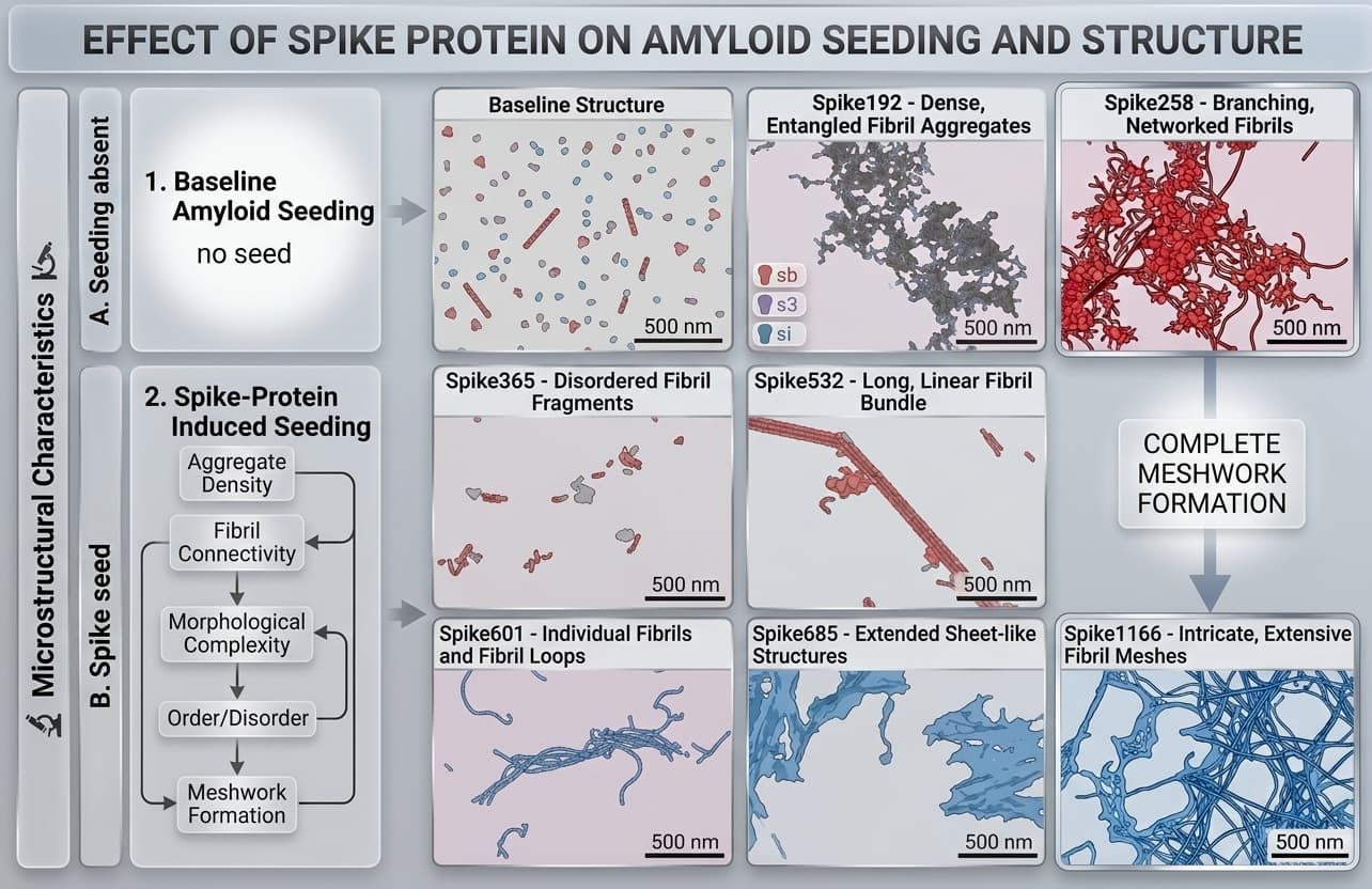
Amyloid microclots behave differently because their protein architecture is altered. In landmark 2025 research published in Biochemistry, Hanna Westman, Per Hammarström, and Sofie Nyström tested seven spike-derived amyloid peptides against purified human clotting factors (Westman et al., 2025). They found that specific spike amyloid sequences, particularly Spike685, incorporate directly into polymerizing fibrin networks and render the resulting clots impervious to plasmin digestion, as covered in spike protein amyloid fibrils and fibrinolysis.
Proteomic analysis of patient microclots confirms this structural resistance. Resia Pretorius, Douglas Kell, and colleagues discovered that circulating microclots trap massive amounts of alpha-2 antiplasmin, the body's primary fibrinolysis inhibitor, alongside von Willebrand factor and platelet factor 4 (Pretorius et al., 2022). The microclot effectively armors itself against enzymatic clearance, allowing it to persist in cerebral capillaries for months or years.
Why standard medical tests miss microclots in brain tissue
Patients suffering from profound post-COVID cognitive decline frequently receive normal results on brain imaging and routine blood panels. This creates severe medical gaslighting, leaving patients told their symptoms are psychosomatic.
The discrepancy stems from diagnostic scale:
| Diagnostic Tool | Target Analyte | Spatial Resolution / Detection Limit | Can It Detect Microclots in Brain? |
|---|---|---|---|
| Head CT Scan | Macroscopic tissue density and acute hemorrhage | 1 to 2 millimeters | No. Misses microscopic capillary obstructions entirely. |
| Brain MRI (1.5T / 3T) | White matter hyperintensities and macroscopic infarcts | 0.5 to 1 millimeter | No. Cannot resolve 5 to 10 µm capillary occlusions. |
| Amyloid PET Scan | Extracellular fibrillar brain parenchymal plaques | 3 to 5 millimeters | No. Measures brain amyloid-beta plaques, not vascular fibrin clots. |
| Routine D-Dimer Test | Soluble fragments of dissolved fibrin clots | Micrograms per milliliter | No. Fails because amyloid microclots resist enzymatic dissolution. |
| ThT Fluorescence Microscopy | Cross-beta sheet amyloid structures in capillary blood | Nanometer fluorophore binding | Yes. Directly visualizes circulating fibrin amyloid microclot burden. |
A normal CT or MRI confirms you haven't suffered a major stroke, but it cannot rule out diffuse microvascular capillary occlusion.
How to test for circulating amyloid microclots
Because standard hospital analyzers cannot see intact microclots, research teams use fluorescence microscopy with Thioflavin T (ThT). ThT is a benzothiazole dye that remains non-fluorescent in free solution. When ThT molecules insert into the cross-beta sheet grooves characteristic of an amyloid fibril, their molecular rotation is sterically locked, producing a 50- to 1,000-fold jump in green fluorescence under blue excitation light.
Our at-home amyloid test uses this proven staining method on a finger-prick dried blood sample, returning a Videographic Microscopy Report that classifies microclot burden across standardized stages. It is an educational screening tool, not a clinical diagnosis of dementia or Alzheimer's disease, and does not measure spike protein directly. It gives patients and their physicians objective observational data on circulating microvascular clot burden.
To compare diagnostic protocols and understand the methodology, read our overview on how to test for microclots and our technical guide on the amyloid fibrin test.
Emerging treatments under active investigation
Because amyloid microclots resist conventional plasmin breakdown, standard blood thinners alone rarely resolve chronic symptoms. Researchers and specialty clinics are evaluating multi-modal therapies designed to eliminate or disassemble these resistant matrices:
- Double Filtration Plasmapheresis (DFPP): At specialized facilities like Edogawa Hospital in Tokyo, Japan, clinicians use two-stage membrane filtration to physically remove high-molecular-weight fibrinogen, autoantibodies, and microclots from plasma, followed by dental pulp stem cell growth factors to support endothelial recovery (plasmapheresis for Long COVID).
- Low-Frequency Acoustic Disruption (150 kHz): Microfluidic lab-on-a-chip studies demonstrate that 150 kHz ultrasound mechanically breaks apart patient-derived amyloid microclots, reducing mean clot diameter by over 60 percent without harming red blood cells (nattokinase and lumbrokinase for microclots).
- Systemic Fibrinolytic Enzymes: Oral proteolytic enzymes like lumbrokinase and serrapeptase are being investigated for their ability to cleave alternative peptide bonds that plasmin cannot access.
Never begin or discontinue anticoagulant or fibrinolytic protocols without direct medical supervision. If you are seeking a physician experienced in post-viral microvascular medicine, explore our doctor directory.
Where to go next
To explore the clinical evidence on capillary hypoperfusion, read our detailed guide on microclots symptoms and capillary hypoperfusion. If you want to understand the laboratory evidence showing how viral proteins initiate cross-seeding in neurodegenerative diseases, review spike amyloid cross-seeding in Alzheimer's. When you are ready to evaluate your own microvascular sample, begin with how to test for microclots.
Frequently asked questions
Can microclots in brain tissue cause dementia or Alzheimer's?
Microclots can starve cerebral tissue of oxygen by blocking capillaries, accelerating cognitive decline and vascular dementia. In laboratory studies, amyloid fibrils derived from the spike protein also directly hetero-seed amyloid-beta and tau, the hallmark proteins of Alzheimer's disease.
Why don't standard brain scans show microclots?
Standard hospital head CT scans and 1.5T MRIs are designed to visualize macroscopic tissue damage, such as large ischemic strokes or bleeding larger than 1 to 2 millimeters. Microclots are microscopic aggregates (2 to 200 micrometers) trapped inside individual capillaries, which standard clinical imaging resolutions cannot resolve.
How does COVID-19 increase long-term dementia risk?
A massive 18.3-million-patient study from England showed that COVID-19 elevates dementia and cognitive impairment risk for up to 4 years. Researchers link this to persistent microvascular hypoperfusion, chronic endothelial inflammation, and cross-beta sheet amyloid fibril deposition that resists normal enzymatic breakdown.
How can you test for microclots in the bloodstream?
Conventional clotting panels like D-dimer miss these particles because they only measure soluble breakdown products. Microclots are detected using Thioflavin T fluorescence microscopy, which binds specifically to cross-beta sheet amyloid structures in capillary or venous blood.
Sources
- Association of COVID-19 with neurodegenerative and neurological disorders in vaccinated and unvaccinated people. medRxiv, 2026
- Amyloidogenesis of SARS-CoV-2 Spike Protein. Journal of the American Chemical Society, 2022
- SARS-CoV-2 Spike Amyloid Fibrils Alter Fibrin Polymerization and Inhibit Fibrinolysis. Biochemistry, 2025
- Proteomics of fibrin amyloid microclots in long COVID/PASC shows many entrapped pro-inflammatory molecules that may also contribute to a failed fibrinolytic system. Cardiovascular Diabetology, 2022
- Long COVID: major findings, mechanisms and recommendations. Nature Reviews Microbiology, 2023
At-Home Amyloid Test
Experiencing persistent brain fog or cognitive fatigue?
Our at-home amyloid test screens your capillary blood for circulating fibrin amyloid microclots using Thioflavin T fluorescence microscopy.
Next steps
- Spike Protein and Alzheimer's: The Spike Amyloid Cross-Seeding StudySpike protein amyloid sped up misfolding of Alzheimer's amyloid-beta and prion protein in a lab study. What it shows about the brain, and what it doesn't.
- Types of Amyloid Tests: Blood Tests, PET, CSF, Biopsy and MicroclotsCompare the types of amyloid tests: Alzheimer's blood tests, amyloid PET, spinal fluid, amyloidosis biopsy and fibrin amyloid microclot microscopy.
- What Are Microclots? Symptoms, Tests and RisksMicroclots are microscopic blood clots studied in Long COVID. Learn what they are, how micro clotting forms, symptoms, tests and risks.

Tim Ottowitz
Founder & Research Lead
Tim Ottowitz founded Amyloid Test and writes its Science Hub, turning fibrin amyloid and spike protein research into plain-language guides that stay close to the published evidence.
Keep reading
Amyloid RNA Delivery: How Seminal Fibrils Shuttle Genetic Cargo
Columbia scientists discovered seminal amyloids shuttle RNA into sperm. Learn how amyloid RNA delivery works and its promise for non-viral gene therapy.
Is Spike Protein a Prion? What a 2026 Prion-Like Domain Review Shows
Is spike protein a prion? A 2026 review links spike prion-like domains to amyloid-beta and tau cross-seeding. Here's what's proven and what isn't yet.
Micro Blood Clots Symptoms: Microclots Symptoms Guide
Micro blood clots symptoms include fatigue, brain fog and breathlessness. Learn why microclots may cause capillary hypoperfusion and what tests can show.